3E4Y

Crystal structure of a 33kDa catalase-related protein from Mycobacterium avium subsp. paratuberculosis. I2(1)2(1)2(1) crystal form


Experimental Data Snapshot

  • Method: X-RAY DIFFRACTION
  • Resolution: 2.60 Å
  • R-Value Free: 0.257 
  • R-Value Work: 0.202 

Starting Model: experimental
View more details

wwPDB Validation   3D Report Full Report


Ligand Structure Quality Assessment 


This is version 1.2 of the entry. See complete history


Literature

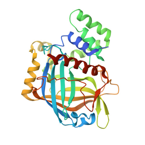
The structure and peroxidase activity of a 33-kDa catalase-related protein from Mycobacterium avium ssp. paratuberculosis.

Pakhomova, S.Gao, B.Boeglin, W.E.Brash, A.R.Newcomer, M.E.

(2009) Protein Sci 18: 2559-2568

  • DOI: https://doi.org/10.1002/pro.265
  • Primary Citation of Related Structures:  
    3E4W, 3E4Y

  • PubMed Abstract: 

    True catalases are tyrosine-liganded, usually tetrameric, hemoproteins with subunit sizes of approximately 55-84 kDa. Recently characterized hemoproteins with a catalase-related structure, yet lacking in catalatic activity, include the 40-43 kDa allene oxide synthases of marine invertebrates and cyanobacteria. Herein, we describe the 1.8 A X-ray crystal structure of a 33 kDa subunit hemoprotein from Mycobacterium avium ssp. paratuberculosis (annotated as MAP-2744c), that retains the core elements of the catalase fold and exhibits an organic peroxide-dependent peroxidase activity. MAP-2744c exhibits negligible catalatic activity, weak peroxidatic activity using hydrogen peroxide (20/s) and strong peroxidase activity (approximately 300/s) using organic hydroperoxides as co-substrate. Key amino acid differences significantly impact prosthetic group conformation and placement and confer a distinct activity to this prototypical member of a group of conserved bacterial "minicatalases". Its structural features and the result of the enzyme assays support a role for MAP-2744c and its close homologues in mitigating challenge by a variety of reactive oxygen species.


  • Organizational Affiliation

    Department of Biological Sciences, Louisiana State University, Baton Rouge, Louisiana 70803, USA.


Macromolecules
Find similar proteins by:  (by identity cutoff)  |  3D Structure
Entity ID: 1
MoleculeChains Sequence LengthOrganismDetailsImage
Putative uncharacterized protein320Mycobacterium avium subsp. paratuberculosisMutation(s): 0 
Gene Names: MAP_2744c
UniProt
Find proteins for Q73WB6 (Mycolicibacterium paratuberculosis (strain ATCC BAA-968 / K-10))
Explore Q73WB6 
Go to UniProtKB:  Q73WB6
Entity Groups  
Sequence Clusters30% Identity50% Identity70% Identity90% Identity95% Identity100% Identity
UniProt GroupQ73WB6
Sequence Annotations
Expand
  • Reference Sequence
Experimental Data & Validation

Experimental Data

  • Method: X-RAY DIFFRACTION
  • Resolution: 2.60 Å
  • R-Value Free: 0.257 
  • R-Value Work: 0.202 
  • Space Group: I 21 21 21
Unit Cell:
Length ( Å )Angle ( ˚ )
a = 69.604α = 90
b = 92.68β = 90
c = 165.499γ = 90
Software Package:
Software NamePurpose
MAR345dtbdata collection
AMoREphasing
CNSrefinement
HKL-2000data reduction
HKL-2000data scaling

Structure Validation

View Full Validation Report


Ligand Structure Quality Assessment 


Entry History 

Deposition Data

Revision History  (Full details and data files)

  • Version 1.0: 2009-08-18
    Type: Initial release
  • Version 1.1: 2011-07-13
    Changes: Non-polymer description, Version format compliance
  • Version 1.2: 2023-08-30
    Changes: Data collection, Database references, Derived calculations, Refinement description