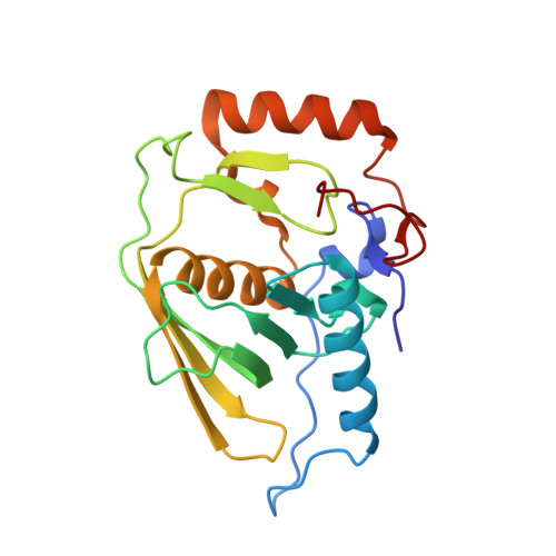
3E3U

Crystal structure of Mycobacterium tuberculosis peptide deformylase in complex with inhibitor


Experimental Data Snapshot

  • Method: X-RAY DIFFRACTION
  • Resolution: 1.56 Å
  • R-Value Free: 0.203 
  • R-Value Work: 0.171 
  • R-Value Observed: 0.173 

wwPDB Validation   3D Report Full Report


Ligand Structure Quality Assessment 


This is version 1.2 of the entry. See complete history


Literature

Peptide deformylase inhibitors of Mycobacterium tuberculosis: synthesis, structural investigations, and biological results.

Pichota, A.Duraiswamy, J.Yin, Z.Keller, T.H.Alam, J.Liung, S.Lee, G.Ding, M.Wang, G.Chan, W.L.Schreiber, M.Ma, I.Beer, D.Ngew, X.Mukherjee, K.Nanjundappa, M.Teo, J.W.Thayalan, P.Yap, A.Dick, T.Meng, W.Xu, M.Koehn, J.Pan, S.H.Clark, K.Xie, X.Shoen, C.Cynamon, M.

(2008) Bioorg Med Chem Lett 18: 6568-6572

  • DOI: https://doi.org/10.1016/j.bmcl.2008.10.040
  • Primary Citation of Related Structures:  
    3E3U

  • PubMed Abstract: 

    Bacterial peptide deformylase (PDF) belongs to a subfamily of metalloproteases catalyzing the removal of the N-terminal formyl group from newly synthesized proteins. We report the synthesis and biological activity of highly potent inhibitors of Mycobacterium tuberculosis (Mtb) PDF enzyme as well as the first X-ray crystal structure of Mtb PDF. Structure-activity relationship and crystallographic data clarified the structural requirements for high enzyme potency and cell based potency. Activities against single and multi-drug-resistant Mtb strains are also reported.


  • Organizational Affiliation

    Novartis Institute for Tropical Diseases, 10 Biopolis Road, #05-01 Chromos, Singapore 138670, Singapore. arkadius.pichota@novartis.com


Macromolecules
Find similar proteins by:  (by identity cutoff)  |  3D Structure
Entity ID: 1
MoleculeChains Sequence LengthOrganismDetailsImage
Peptide deformylase197Mycobacterium tuberculosisMutation(s): 0 
Gene Names: def
EC: 3.5.1.88
UniProt
Find proteins for P9WIJ3 (Mycobacterium tuberculosis (strain ATCC 25618 / H37Rv))
Explore P9WIJ3 
Go to UniProtKB:  P9WIJ3
Entity Groups  
Sequence Clusters30% Identity50% Identity70% Identity90% Identity95% Identity100% Identity
UniProt GroupP9WIJ3
Sequence Annotations
Expand
  • Reference Sequence
Small Molecules
Binding Affinity Annotations 
IDSourceBinding Affinity
NVC PDBBind:  3E3U IC50: 13 (nM) from 1 assay(s)
BindingDB:  3E3U IC50: 13 (nM) from 1 assay(s)
Experimental Data & Validation

Experimental Data

  • Method: X-RAY DIFFRACTION
  • Resolution: 1.56 Å
  • R-Value Free: 0.203 
  • R-Value Work: 0.171 
  • R-Value Observed: 0.173 
  • Space Group: C 1 2 1
Unit Cell:
Length ( Å )Angle ( ˚ )
a = 100.87α = 90
b = 54.765β = 94.39
c = 40.215γ = 90
Software Package:
Software NamePurpose
CNSrefinement
PDB_EXTRACTdata extraction
HKL-2000data collection
HKL-2000data reduction
HKL-2000data scaling
REFMACphasing
REFMACrefinement

Structure Validation

View Full Validation Report


Ligand Structure Quality Assessment 


Entry History 

Deposition Data

Revision History  (Full details and data files)

  • Version 1.0: 2009-01-20
    Type: Initial release
  • Version 1.1: 2011-07-13
    Changes: Version format compliance
  • Version 1.2: 2024-02-21
    Changes: Data collection, Database references, Derived calculations