3DWK | pdb_00003dwk

Identification of Dynamic Structural Motifs Involved in Peptidoglycan Glycosyltransfer


Experimental Data Snapshot

  • Method: X-RAY DIFFRACTION
  • Resolution: 3.10 Å
  • R-Value Free: 
    0.264 (Depositor), 0.243 (DCC) 
  • R-Value Work: 
    0.218 (Depositor) 
  • R-Value Observed: 
    0.218 (Depositor) 

Starting Model: experimental
View more details

wwPDB Validation 3D Report Full Report

Validation slider image for 3DWK

Ligand Structure Quality Assessment 


This is version 1.3 of the entry. See complete history

Literature

Identification of dynamic structural motifs involved in peptidoglycan glycosyltransfer.

Lovering, A.L.De Castro, L.Strynadka, N.C.

(2008) J Mol Biology 383: 167-177

  • DOI: https://doi.org/10.1016/j.jmb.2008.08.020
  • Primary Citation Related Structures: 
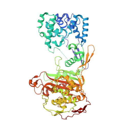
    3DWK

  • PubMed Abstract: 

    We have determined the structure of a new form of the bifunctional peptidoglycan glycosyltransferase (GT)/transpeptidase penicillin-binding protein 2 from the pathogen Staphylococcus aureus. We observe several previously unstructured regions of the GT substrate-binding pockets, including a pi-bulge in the outer helix that may be responsible for the conformational flexibility of active-site motifs required for transfer of product to the donor binding site during processive rounds of peptidoglycan polymerization. The identification of a beta-hairpin in the usually unstructured region of the fold shares local structural homology to that of an exomuramidase, heightening comparisons between this biosynthetic enzyme and lytic peptidoglycan transglycosylases. This new form also shows remarkable interdomain flexibility, causing the linker region of the fold to project into the GT active site. This self-interaction may have significant consequences for the regulation of polymerization activity. The derived information is used to build a catalytic model of both donor and acceptor glycolipid substrates.


  • Organizational Affiliation
    • Department of Biochemistry and Molecular Biology, University of British Columbia, Vancouver, Canada.

Macromolecule Content 

  • Total Structure Weight: 283.16 kDa 
  • Atom Count: 19,474 
  • Modeled Residue Count: 2,444 
  • Deposited Residue Count: 2,500 
  • Unique protein chains: 1

Macromolecules

Find similar proteins by:|  3D Structure
Entity ID: 1
MoleculeChains  Sequence LengthOrganismDetailsImage
Penicillin-binding protein 2
A, B, C, D
625Staphylococcus aureus subsp. aureus COLMutation(s): 1 
Gene Names: pbp2SACOL1490
UniProt
Find proteins for Q8KHY3 (Staphylococcus aureus)
Explore Q8KHY3 
Go to UniProtKB:  Q8KHY3
Entity Groups
Sequence Clusters30% Identity50% Identity70% Identity90% Identity95% Identity100% Identity
UniProt GroupQ8KHY3
Sequence Annotations
Expand
Reference Sequence

Small Molecules

Ligands 2 Unique
IDChains Name / Formula / InChI Key2D Diagram3D Interactions
LDA

Query on LDA


Download:Ideal Coordinates CCD File
YA [auth D]LAURYL DIMETHYLAMINE-N-OXIDE
C14 H31 N O
SYELZBGXAIXKHU-UHFFFAOYSA-N
SO4

Query on SO4


Download:Ideal Coordinates CCD File
AA [auth B]
BA [auth B]
CA [auth B]
DA [auth B]
E [auth A]
AA [auth B],
BA [auth B],
CA [auth B],
DA [auth B],
E [auth A],
EA [auth B],
F [auth A],
FA [auth B],
G [auth A],
GA [auth C],
H [auth A],
HA [auth C],
I [auth A],
IA [auth C],
J [auth A],
JA [auth C],
K [auth A],
KA [auth C],
L [auth A],
LA [auth C],
M [auth A],
MA [auth C],
N [auth A],
NA [auth C],
O [auth A],
OA [auth C],
P [auth A],
PA [auth D],
Q [auth A],
QA [auth D],
R [auth A],
RA [auth D],
S [auth A],
SA [auth D],
T [auth B],
TA [auth D],
U [auth B],
UA [auth D],
V [auth B],
VA [auth D],
W [auth B],
WA [auth D],
X [auth B],
XA [auth D],
Y [auth B],
Z [auth B]
SULFATE ION
O4 S
QAOWNCQODCNURD-UHFFFAOYSA-L

Experimental Data & Validation

Experimental Data

  • Method: X-RAY DIFFRACTION
  • Resolution: 3.10 Å
  • R-Value Free:  0.264 (Depositor), 0.243 (DCC) 
  • R-Value Work:  0.218 (Depositor) 
  • R-Value Observed: 0.218 (Depositor) 
Space Group: P 1 21 1
Unit Cell:
Length ( Å )Angle ( ˚ )
a = 85.352α = 90
b = 217.471β = 90.3
c = 95.735γ = 90
Software Package:
Software NamePurpose
MOSFLMdata reduction
SCALAdata scaling
PHASERphasing
REFMACrefinement
PDB_EXTRACTdata extraction
ADSCdata collection

Structure Validation

View Full Validation Report


Ligand Structure Quality Assessment 


Entry History 

Deposition Data

Revision History  (Full details and data files)

  • Version 1.0: 2008-09-30
    Type: Initial release
  • Version 1.1: 2011-07-13
    Changes: Version format compliance
  • Version 1.2: 2021-10-20
    Changes: Database references, Derived calculations
  • Version 1.3: 2023-08-30
    Changes: Data collection, Refinement description