3DOF

Complex of ARL2 and BART, Crystal Form 2


Experimental Data Snapshot

  • Method: X-RAY DIFFRACTION
  • Resolution: 3.30 Å
  • R-Value Free: 0.294 
  • R-Value Work: 0.253 
  • R-Value Observed: 0.255 

wwPDB Validation   3D Report Full Report


Ligand Structure Quality Assessment 


This is version 1.2 of the entry. See complete history


Literature

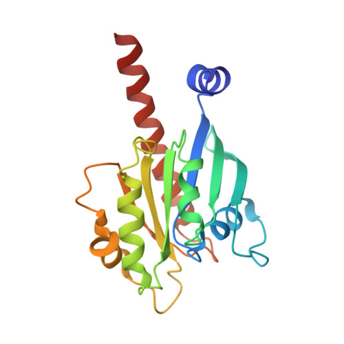
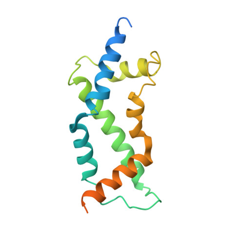
Crystal structure of the ARL2-GTP-BART complex reveals a novel recognition and binding mode of small GTPase with effector

Zhang, T.Li, S.Zhang, Y.Zhong, C.Lai, Z.Ding, J.

(2009) Structure 17: 602-610

  • DOI: https://doi.org/10.1016/j.str.2009.01.014
  • Primary Citation of Related Structures:  
    3DOE, 3DOF

  • PubMed Abstract: 

    ARL2 is a member of the ADP-ribosylation factor family but has unique biochemical features. BART is an effector of ARL2 that is essential for nuclear retention of STAT3 and may also be involved in mitochondria transport and apoptosis. Here we report the crystal structure and biochemical characterization of human ARL2-GTP-BART complex. ARL2-GTP assumes a typical small GTPase fold with a unique N-terminal alpha helix conformation. BART consists of a six alpha helix bundle. The interactions between ARL2 and BART involve two interfaces: a conserved N-terminal LLXIL motif of ARL2 is embedded in a hydrophobic cleft of BART and the switch regions of ARL2 interact with helix alpha3 of BART. Both interfaces are essential for the binding as verified by mutagenesis study. This novel recognition and binding mode is different from that of other small GTPase-effector interactions and provides molecular basis for the high specificity of ARL2 for BART.


  • Organizational Affiliation

    Institute of Biochemistry and Cell Biology, Shanghai Institutes for Biological Sciences, Shanghai 200031, China.


Macromolecules
Find similar proteins by:  (by identity cutoff)  |  3D Structure
Entity ID: 1
MoleculeChains Sequence LengthOrganismDetailsImage
ADP-ribosylation factor-like protein 2192Homo sapiensMutation(s): 0 
UniProt & NIH Common Fund Data Resources
Find proteins for P36404 (Homo sapiens)
Explore P36404 
Go to UniProtKB:  P36404
PHAROS:  P36404
GTEx:  ENSG00000213465 
Entity Groups  
Sequence Clusters30% Identity50% Identity70% Identity90% Identity95% Identity100% Identity
UniProt GroupP36404
Sequence Annotations
Expand
  • Reference Sequence
Find similar proteins by:  (by identity cutoff)  |  3D Structure
Entity ID: 2
MoleculeChains Sequence LengthOrganismDetailsImage
ADP-ribosylation factor-like protein 2-binding protein165Homo sapiensMutation(s): 0 
UniProt & NIH Common Fund Data Resources
Find proteins for Q9Y2Y0 (Homo sapiens)
Explore Q9Y2Y0 
Go to UniProtKB:  Q9Y2Y0
PHAROS:  Q9Y2Y0
GTEx:  ENSG00000102931 
Entity Groups  
Sequence Clusters30% Identity50% Identity70% Identity90% Identity95% Identity100% Identity
UniProt GroupQ9Y2Y0
Sequence Annotations
Expand
  • Reference Sequence
Small Molecules
Ligands 2 Unique
IDChains Name / Formula / InChI Key2D Diagram3D Interactions
GTP
Query on GTP

Download Ideal Coordinates CCD File 
C [auth A]GUANOSINE-5'-TRIPHOSPHATE
C10 H16 N5 O14 P3
XKMLYUALXHKNFT-UUOKFMHZSA-N
MG
Query on MG

Download Ideal Coordinates CCD File 
D [auth A]MAGNESIUM ION
Mg
JLVVSXFLKOJNIY-UHFFFAOYSA-N
Experimental Data & Validation

Experimental Data

  • Method: X-RAY DIFFRACTION
  • Resolution: 3.30 Å
  • R-Value Free: 0.294 
  • R-Value Work: 0.253 
  • R-Value Observed: 0.255 
  • Space Group: P 21 21 21
Unit Cell:
Length ( Å )Angle ( ˚ )
a = 44.663α = 90
b = 45.165β = 90
c = 175.879γ = 90
Software Package:
Software NamePurpose
REFMACrefinement
HKL-2000data collection
HKL-2000data reduction
HKL-2000data scaling
CNSphasing

Structure Validation

View Full Validation Report


Ligand Structure Quality Assessment 


Entry History 

Deposition Data

Revision History  (Full details and data files)

  • Version 1.0: 2009-03-03
    Type: Initial release
  • Version 1.1: 2011-07-13
    Changes: Version format compliance
  • Version 1.2: 2023-11-01
    Changes: Data collection, Database references, Derived calculations, Refinement description