


Crystal structure of mutant ABL kinase domain in complex with small molecule fragment
Bounaud, P.-Y., Gosberg, A., Hendle, J., Lewis, H.A., Romero, R., Wilson, M.E., Zhang, F.To be published.
Experimental Data Snapshot
Entity ID: 1 | |||||
|---|---|---|---|---|---|
| Molecule | Chains | Sequence Length | Organism | Details | Image |
| Proto-oncogene tyrosine-protein kinase ABL1 | 293 | Mus musculus | Mutation(s): 2 Gene Names: Abl1, Abl EC: 2.7.10.2 | 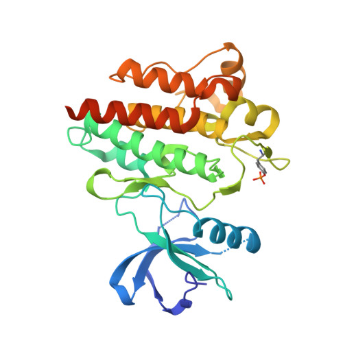 | |
UniProt | |||||
Entity Groups | |||||
| Sequence Clusters | 30% Identity50% Identity70% Identity90% Identity95% Identity100% Identity | ||||
| UniProt Group | P00520 | ||||
Sequence AnnotationsExpand | |||||
Reference Sequence | |||||
| Ligands 1 Unique | |||||
|---|---|---|---|---|---|
| ID | Chains | Name / Formula / InChI Key | 2D Diagram | 3D Interactions | |
| SX7 Download:Ideal Coordinates CCD File | C [auth A], D [auth B] | 2-amino-5-[3-(1-ethyl-1H-pyrazol-5-yl)-1H-pyrrolo[2,3-b]pyridin-5-yl]-N,N-dimethylbenzamide C21 H22 N6 O INAGORZAOFUKOZ-UHFFFAOYSA-N |  | ||
| Modified Residues 1 Unique | |||||
|---|---|---|---|---|---|
| ID | Chains | Type | Formula | 2D Diagram | Parent |
| PTR Query on PTR | A, B | L-PEPTIDE LINKING | C9 H12 N O6 P |  | TYR |
| Length ( Å ) | Angle ( ˚ ) |
|---|---|
| a = 105.356 | α = 90 |
| b = 131.908 | β = 90 |
| c = 56.641 | γ = 90 |
| Software Name | Purpose |
|---|---|
| SCALA | data scaling |
| REFMAC | refinement |
| PDB_EXTRACT | data extraction |


