


Crystal Structure of apo form (Zinc removed) of the Botulinum Neurotoxin Type C Light Chain
Rawat, R., Kumaran, D., Swaminathan, S.To be published.
Experimental Data Snapshot
Starting Model: experimental
View more details
wwPDB Validation 3D Report Full Report

Entity ID: 1 | |||||
|---|---|---|---|---|---|
| Molecule | Chains | Sequence Length | Organism | Details | Image |
| Botulinum neurotoxin C1 light chain | 450 | Clostridium botulinum | Mutation(s): 0 Gene Names: BoNT/C EC: 3.4.24.69 | 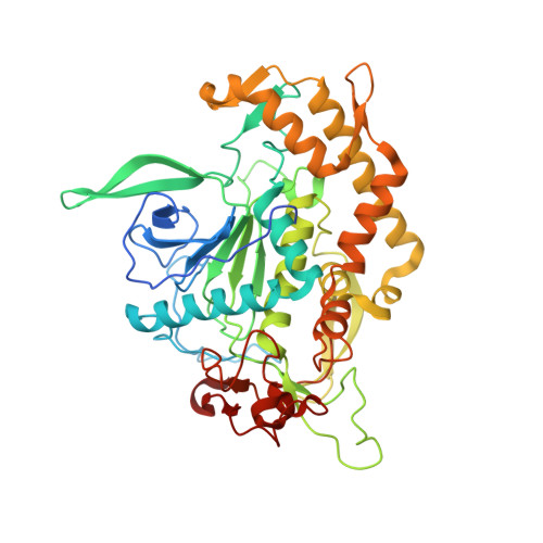 | |
UniProt | |||||
Entity Groups | |||||
| Sequence Clusters | 30% Identity50% Identity70% Identity90% Identity95% Identity100% Identity | ||||
| UniProt Group | P18640 | ||||
Sequence AnnotationsExpand | |||||
Reference Sequence | |||||
| Ligands 4 Unique | |||||
|---|---|---|---|---|---|
| ID | Chains | Name / Formula / InChI Key | 2D Diagram | 3D Interactions | |
| ACT Download:Ideal Coordinates CCD File | D [auth A] | ACETATE ION C2 H3 O2 QTBSBXVTEAMEQO-UHFFFAOYSA-M |  | ||
| FMT Download:Ideal Coordinates CCD File | E [auth A] F [auth A] G [auth A] H [auth A] I [auth A] | FORMIC ACID C H2 O2 BDAGIHXWWSANSR-UHFFFAOYSA-N |  | ||
| CA Download:Ideal Coordinates CCD File | C [auth A] | CALCIUM ION Ca BHPQYMZQTOCNFJ-UHFFFAOYSA-N |  | ||
| NA Download:Ideal Coordinates CCD File | B [auth A] | SODIUM ION Na FKNQFGJONOIPTF-UHFFFAOYSA-N |  | ||
| Length ( Å ) | Angle ( ˚ ) |
|---|---|
| a = 106.74 | α = 90 |
| b = 106.74 | β = 90 |
| c = 140.24 | γ = 120 |
| Software Name | Purpose |
|---|---|
| CNS | refinement |
| CBASS | data collection |
| HKL-2000 | data reduction |
| HKL-2000 | data scaling |
| MOLREP | phasing |


