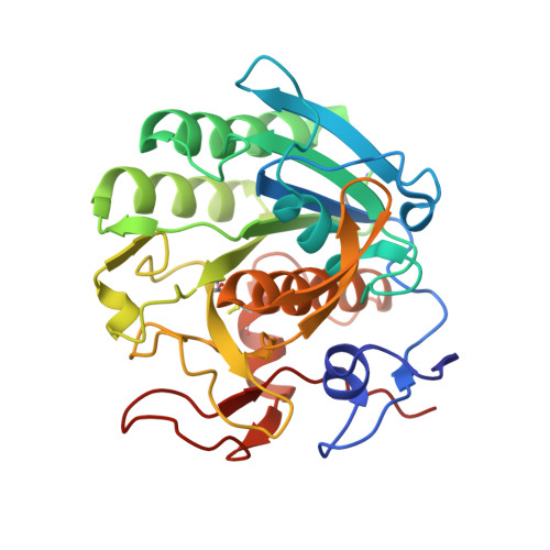
3DE7

Proteinase K by Classical hanging drop method after the fourth step of high X-Ray dose on ESRF ID23-1 beamline


Experimental Data Snapshot

  • Method: X-RAY DIFFRACTION
  • Resolution: 2.30 Å
  • R-Value Free: 0.211 
  • R-Value Work: 0.161 
  • R-Value Observed: 0.163 

wwPDB Validation   3D Report Full Report


This is version 1.1 of the entry. See complete history


Literature

Radiation stability of proteinase K crystals grown by LB nanotemplate method

Pechkova, E.Tripathi, S.Ravelli, R.B.McSweeney, S.Nicolini, C.

(2009) J Struct Biol 168: 409-418

  • DOI: https://doi.org/10.1016/j.jsb.2009.08.005
  • Primary Citation of Related Structures:  
    3D9Q, 3DDZ, 3DE0, 3DE1, 3DE2, 3DE3, 3DE4, 3DE5, 3DE6, 3DE7

  • PubMed Abstract: 

    A detailed analysis of structural and intensity changes induced by X-ray radiation is presented for two types of proteinase K crystals: crystal grown by classical hanging drop method and those grown by Langmuir-Blodgett (LB) nanotemplate. The comparison of various parameters (e.g. intensity per sigma ratio, unit-cell volume, number of unique reflections, B-factors) and electron density maps as a function of radiation dose, demonstrates that crystals, grown by the LB nanotemplate method, appear to be more resistant against radiation damage than crystals grown by the classical hanging drop method.


  • Organizational Affiliation

    Nanoworld Institute, CIRSDNNOB University of Genova, Genoa, Italy.


Macromolecules
Find similar proteins by:  (by identity cutoff)  |  3D Structure
Entity ID: 1
MoleculeChains Sequence LengthOrganismDetailsImage
Proteinase KA [auth X]279Parengyodontium albumMutation(s): 0 
EC: 3.4.21.64
UniProt
Find proteins for P06873 (Parengyodontium album)
Explore P06873 
Go to UniProtKB:  P06873
Entity Groups  
Sequence Clusters30% Identity50% Identity70% Identity90% Identity95% Identity100% Identity
UniProt GroupP06873
Sequence Annotations
Expand
  • Reference Sequence
Small Molecules
Ligands 1 Unique
IDChains Name / Formula / InChI Key2D Diagram3D Interactions
CA
Query on CA

Download Ideal Coordinates CCD File 
B [auth X]CALCIUM ION
Ca
BHPQYMZQTOCNFJ-UHFFFAOYSA-N
Experimental Data & Validation

Experimental Data

  • Method: X-RAY DIFFRACTION
  • Resolution: 2.30 Å
  • R-Value Free: 0.211 
  • R-Value Work: 0.161 
  • R-Value Observed: 0.163 
  • Space Group: P 43 21 2
Unit Cell:
Length ( Å )Angle ( ˚ )
a = 67.963α = 90
b = 67.963β = 90
c = 102.809γ = 90
Software Package:
Software NamePurpose
MOSFLMdata reduction
SCALAdata scaling
REFMACrefinement
PDB_EXTRACTdata extraction
ADSCdata collection

Structure Validation

View Full Validation Report


Entry History 

Deposition Data

Revision History  (Full details and data files)

  • Version 1.0: 2009-06-09
    Type: Initial release
  • Version 1.1: 2011-07-13
    Changes: Version format compliance