3D6Y | pdb_00003d6y

Crystal structure of R275E mutant of BMRR bound to DNA and berberine


Experimental Data Snapshot

  • Method: X-RAY DIFFRACTION
  • Resolution: 2.70 Å
  • R-Value Free: 
    0.259 (Depositor) 
  • R-Value Work: 
    0.227 (Depositor), 0.231 (DCC) 
  • R-Value Observed: 
    0.227 (Depositor) 

Starting Model: experimental
View more details

wwPDB Validation 3D Report Full Report

Validation slider image for 3D6Y

Ligand Structure Quality Assessment 


This is version 1.3 of the entry. See complete history

Literature

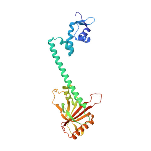
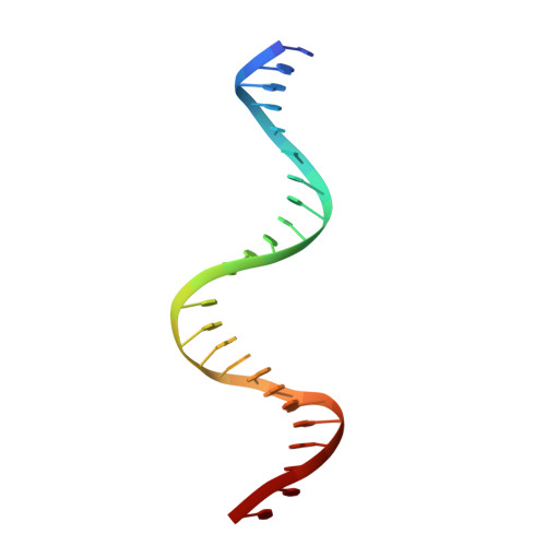
Structures of BmrR-Drug Complexes Reveal a Rigid Multidrug Binding Pocket and Transcription Activation through Tyrosine Expulsion

Newberry, K.J.Huffman, J.L.Miller, M.C.Vazquez-Laslop, N.Neyfakh, A.A.Brennan, R.G.

(2008) J Biological Chem 283: 26795-26804

  • DOI: https://doi.org/10.1074/jbc.M804191200
  • Primary Citation Related Structures: 
    3D6Y, 3D6Z, 3D70, 3D71

  • PubMed Abstract: 

    BmrR is a member of the MerR family and a multidrug binding transcription factor that up-regulates the expression of the bmr multidrug efflux transporter gene in response to myriad lipophilic cationic compounds. The structural mechanism by which BmrR binds these chemically and structurally different drugs and subsequently activates transcription is poorly understood. Here, we describe the crystal structures of BmrR bound to rhodamine 6G (R6G) or berberine (Ber) and cognate DNA. These structures reveal each drug stacks against multiple aromatic residues with their positive charges most proximal to the carboxylate group of Glu-253 and that, unlike other multidrug binding pockets, that of BmrR is rigid. Substitution of Glu-253 with either alanine (E253A) or glutamine (E253Q) results in unpredictable binding affinities for R6G, Ber, and tetraphenylphosphonium. Moreover, these drug binding studies reveal that the negative charge of Glu-253 is not important for high affinity binding to Ber and tetraphenylphosphonium but plays a more significant, but unpredictable, role in R6G binding. In vitro transcription data show that E253A and E253Q are constitutively active, and structures of the drug-free E253A-DNA and E253Q-DNA complexes support a transcription activation mechanism requiring the expulsion of Tyr-152 from the multidrug binding pocket. In sum, these data delineate the mechanism by which BmrR binds lipophilic, monovalent cationic compounds and suggest the importance of the redundant negative electrostatic nature of this rigid drug binding pocket that can be used to discriminate against molecules that are not substrates of the Bmr multidrug efflux pump.


  • Organizational Affiliation
    • Department of Biochemistry and Molecular Biology, University of Texas M.D. Anderson Cancer Center, Houston, Texas 77030-4009, USA.

Macromolecule Content 

  • Total Structure Weight: 41.33 kDa 
  • Atom Count: 2,847 
  • Modeled Residue Count: 301 
  • Deposited Residue Count: 308 
  • Unique protein chains: 1
  • Unique nucleic acid chains: 1

Macromolecules


Find similar proteins by:|  3D Structure
Entity ID: 2
MoleculeChains  Sequence LengthOrganismDetailsImage
Multidrug-efflux transporter 1 regulatorB [auth A]284Bacillus subtilisMutation(s): 3 
Gene Names: bmrRbmr1R
UniProt
Find proteins for P39075 (Bacillus subtilis (strain 168))
Explore P39075 
Go to UniProtKB:  P39075
Entity Groups
Sequence Clusters30% Identity50% Identity70% Identity90% Identity95% Identity100% Identity
UniProt GroupP39075
Sequence Annotations
Expand
Reference Sequence
Find similar nucleic acids by:  Sequence
Entity ID: 1
MoleculeChains LengthOrganismImage
BMR promoter DNAA [auth B]24synthetic construct
Sequence Annotations
Expand
Reference Sequence

Experimental Data & Validation

Experimental Data

  • Method: X-RAY DIFFRACTION
  • Resolution: 2.70 Å
  • R-Value Free:  0.259 (Depositor) 
  • R-Value Work:  0.227 (Depositor), 0.231 (DCC) 
  • R-Value Observed: 0.227 (Depositor) 
Space Group: P 43 2 2
Unit Cell:
Length ( Å )Angle ( ˚ )
a = 106.33α = 90
b = 106.33β = 90
c = 146.75γ = 90
Software Package:
Software NamePurpose
CNSrefinement
ADSCdata collection
MOSFLMdata reduction
SCALAdata scaling
EPMRphasing

Structure Validation

View Full Validation Report


Ligand Structure Quality Assessment 


Entry History 

Deposition Data

Revision History  (Full details and data files)

  • Version 1.0: 2008-08-26
    Type: Initial release
  • Version 1.1: 2011-07-13
    Changes: Non-polymer description, Version format compliance
  • Version 1.2: 2021-10-20
    Changes: Database references, Derived calculations, Source and taxonomy
  • Version 1.3: 2023-08-30
    Changes: Data collection, Refinement description