3D5I | pdb_00003d5i

Crystal structure of ribonuclease Sa2 with exo-2',3'-cyclophosphorotioate


Experimental Data Snapshot

  • Method: X-RAY DIFFRACTION
  • Resolution: 2.20 Å
  • R-Value Free: 
    0.262 (Depositor), 0.268 (DCC) 
  • R-Value Work: 
    0.208 (Depositor), 0.212 (DCC) 
  • R-Value Observed: 
    0.210 (Depositor) 

Starting Model: experimental
View more details

wwPDB Validation 3D Report Full Report

Validation slider image for 3D5I

Ligand Structure Quality Assessment 


This is version 1.3 of the entry. See complete history

Literature

Structure of RNase Sa2 complexes with mononucleotides - new aspects of catalytic reaction and substrate recognition

Bauerova-Hlinkova, V.Dvorsky, R.Perecko, D.Povazanec, F.Sevcik, J.

(2009) FEBS J 276: 4156-4168

  • DOI: https://doi.org/10.1111/j.1742-4658.2009.07125.x
  • Primary Citation Related Structures: 
    3D4A, 3D5G, 3D5I, 3DGY, 3DH2

  • PubMed Abstract: 

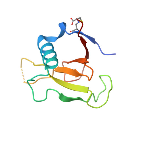
    Although the mechanism of RNA cleavage by RNases has been studied for many years, there remain aspects that have not yet been fully clarified. We have solved the crystal structures of RNase Sa2 in the apo form and in complexes with mononucleotides. These structures provide more details about the mechanism of RNA cleavage by RNase Sa2. In addition to Glu56 and His86, which are the principal catalytic residues, an important role in the first reaction step of RNA cleavage also seems to be played by Arg67 and Arg71, which are located in the phosphate-binding site and form hydrogen bonds with the oxygens of the phosphate group of the mononucleotides. Their positive charge very likely causes polarization of the bonds between the oxygens and the phosphorus atom, leading to electron deficiency on the phosphorus atom and facilitating nucleophilic attack by O2' of the ribose on the phosphorus atom, leading to cyclophosphate formation. The negatively charged Glu56 is in position to attract the proton from O2' of the ribose. Extended molecular docking of mononucleotides, dinucleotides and trinucleotides into the active site of the enzyme allowed us to better understand the guanosine specificity of RNase Sa2 and to predict possible binding subsites for the downstream base and ribose of the second and third nucleotides.


  • Organizational Affiliation
    • Institute of Molecular Biology, Slovak Academy of Sciences, Bratislava, Slovakia. vladena.hlinkova@savba.sk

Macromolecule Content 

  • Total Structure Weight: 33.38 kDa 
  • Atom Count: 2,449 
  • Modeled Residue Count: 279 
  • Deposited Residue Count: 291 
  • Unique protein chains: 1

Macromolecules

Find similar proteins by:|  3D Structure
Entity ID: 1
MoleculeChains  Sequence LengthOrganismDetailsImage
Ribonuclease
A, B, C
97Kitasatospora aureofaciensMutation(s): 0 
EC: 3.1.4.8
UniProt
Find proteins for Q53752 (Kitasatospora aureofaciens)
Explore Q53752 
Go to UniProtKB:  Q53752
Entity Groups
Sequence Clusters30% Identity50% Identity70% Identity90% Identity95% Identity100% Identity
UniProt GroupQ53752
Sequence Annotations
Expand
Reference Sequence

Experimental Data & Validation

Experimental Data

  • Method: X-RAY DIFFRACTION
  • Resolution: 2.20 Å
  • R-Value Free:  0.262 (Depositor), 0.268 (DCC) 
  • R-Value Work:  0.208 (Depositor), 0.212 (DCC) 
  • R-Value Observed: 0.210 (Depositor) 
Space Group: C 1 2 1
Unit Cell:
Length ( Å )Angle ( ˚ )
a = 101.221α = 90
b = 66.775β = 100.8
c = 57.07γ = 90
Software Package:
Software NamePurpose
REFMACrefinement
DENZOdata reduction
SCALEPACKdata scaling
MOLREPphasing

Structure Validation

View Full Validation Report


Ligand Structure Quality Assessment 


Entry History 

Deposition Data

Revision History  (Full details and data files)

  • Version 1.0: 2009-05-26
    Type: Initial release
  • Version 1.1: 2011-07-13
    Changes: Advisory, Version format compliance
  • Version 1.2: 2023-11-01
    Changes: Data collection, Database references, Derived calculations, Refinement description
  • Version 1.3: 2024-10-09
    Changes: Structure summary