3CYE | pdb_00003cye

Cyrstal structure of the native 1918 H1N1 neuraminidase from a crystal with lattice-translocation defects


Experimental Data Snapshot

  • Method: X-RAY DIFFRACTION
  • Resolution: 1.65 Å
  • R-Value Free: 
    0.231 (Depositor), 0.223 (DCC) 
  • R-Value Work: 
    0.193 (Depositor), 0.186 (DCC) 

Starting Model: experimental
View more details

wwPDB Validation 3D Report Full Report

Validation slider image for 3CYE

Ligand Structure Quality Assessment 


This is version 2.2 of the entry. See complete history

Literature

Structure determination of the 1918 H1N1 neuraminidase from a crystal with lattice-translocation defects

Zhu, X.Xu, X.Wilson, I.A.

(2008) Acta Crystallogr D Biol Crystallogr 64: 843-850

  • DOI: https://doi.org/10.1107/S0907444908016648
  • Primary Citation Related Structures: 
    3CYE

  • PubMed Abstract: 

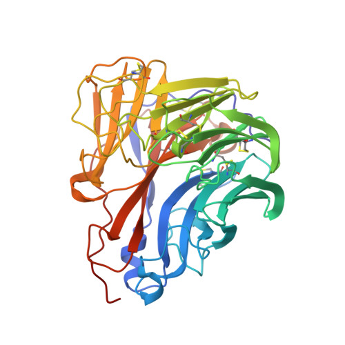
    Few examples of macromolecular crystals containing lattice-translocation defects have been published in the literature. Lattice translocation and twinning are believed to be two common but different crystal-growth anomalies. While the successful use of twinned data for structure determination has become relatively routine in recent years, structure determination of crystals with lattice-translocation defects has not often been reported. To date, only four protein crystal structures containing such a crystal defect have been determined, using corrected, but not uncorrected, intensity data. In this report, the crystallization, structure determination and refinement of N1 neuraminidase derived from the 1918 H1N1 influenza virus (18NA) at 1.65 A resolution are described. The crystal was indexed in space group C222(1), with unit-cell parameters a = 117.7, b = 138.5, c = 117.9 A, and the structure was solved by molecular replacement. The lattice-translocation vector in the 18NA crystal was (0, 1/2, 1/2) or its equivalent vector (1/2, 0, 1/2) owing to the C lattice symmetry. Owing to this special lattice-translocation vector in space group C222(1), structure refinement could be achieved in two different ways: using corrected or uncorrected diffraction data. In the refinement with uncorrected data, a composite model was built to represent the molecules in the translated and untranslated layers, respectively. This composite structure model provided a unique example to examine how the molecules were arranged in the two lattice domains resulting from lattice-translocation defects.


  • Organizational Affiliation
    • Department of Molecular Biology, The Scripps Research Institute, 10550 North Torrey Pines Road, La Jolla, CA 92037, USA.

Macromolecule Content 

  • Total Structure Weight: 87.46 kDa 
  • Atom Count: 6,855 
  • Modeled Residue Count: 770 
  • Deposited Residue Count: 774 
  • Unique protein chains: 1

Macromolecules

Find similar proteins by:|  3D Structure
Entity ID: 1
MoleculeChains  Sequence LengthOrganismDetailsImage
Neuraminidase
A, B
387Influenza A virusMutation(s): 0 
Gene Names: NA
EC: 3.2.1.18
UniProt
Find proteins for Q9IGQ6 (Influenza A virus (strain A/Brevig Mission/1/1918 H1N1))
Explore Q9IGQ6 
Go to UniProtKB:  Q9IGQ6
Entity Groups
Sequence Clusters30% Identity50% Identity70% Identity90% Identity95% Identity100% Identity
UniProt GroupQ9IGQ6
Glycosylation
Glycosylation Sites: 1
Sequence Annotations
Expand
Reference Sequence

Oligosaccharides

Help  
Entity ID: 2
MoleculeChains Length2D Diagram GlycosylationD Interactions
alpha-D-mannopyranose-(1-3)-alpha-D-mannopyranose-(1-4)-2-acetamido-2-deoxy-beta-D-glucopyranose-(1-4)-2-acetamido-2-deoxy-beta-D-glucopyranose
C
4N-Glycosylation
Glycosylation Resources
GlyTouCan: G48068RF
GlyCosmos: G48068RF
GlyGen: G48068RF

Small Molecules

Ligands 5 Unique
IDChains Name / Formula / InChI Key2D Diagram3D Interactions
NAG

Query on NAG


Download:Ideal Coordinates CCD File
D [auth A]2-acetamido-2-deoxy-beta-D-glucopyranose
C8 H15 N O6
OVRNDRQMDRJTHS-FMDGEEDCSA-N
PO4

Query on PO4


Download:Ideal Coordinates CCD File
K [auth A]PHOSPHATE ION
O4 P
NBIIXXVUZAFLBC-UHFFFAOYSA-K
GOL

Query on GOL


Download:Ideal Coordinates CCD File
L [auth A]
M [auth A]
N [auth A]
O [auth A]
P [auth A]
L [auth A],
M [auth A],
N [auth A],
O [auth A],
P [auth A],
U [auth B],
V [auth B],
W [auth B],
X [auth B],
Y [auth B]
GLYCEROL
C3 H8 O3
PEDCQBHIVMGVHV-UHFFFAOYSA-N
ACT

Query on ACT


Download:Ideal Coordinates CCD File
H [auth A],
I [auth A],
J [auth A],
S [auth B],
T [auth B]
ACETATE ION
C2 H3 O2
QTBSBXVTEAMEQO-UHFFFAOYSA-M
CA

Query on CA


Download:Ideal Coordinates CCD File
E [auth A],
F [auth A],
G [auth A],
Q [auth B],
R [auth B]
CALCIUM ION
Ca
BHPQYMZQTOCNFJ-UHFFFAOYSA-N

Experimental Data & Validation

Experimental Data

  • Method: X-RAY DIFFRACTION
  • Resolution: 1.65 Å
  • R-Value Free:  0.231 (Depositor), 0.223 (DCC) 
  • R-Value Work:  0.193 (Depositor), 0.186 (DCC) 
Space Group: C 2 2 21
Unit Cell:
Length ( Å )Angle ( ˚ )
a = 117.728α = 90
b = 138.472β = 90
c = 117.86γ = 90
Software Package:
Software NamePurpose
DENZOdata reduction
SCALEPACKdata scaling
PHASERphasing
REFMACrefinement
PDB_EXTRACTdata extraction
HKL-2000data collection
HKL-2000data reduction
HKL-2000data scaling

Structure Validation

View Full Validation Report


Ligand Structure Quality Assessment 


Entry History 

Deposition Data

Revision History  (Full details and data files)

  • Version 1.0: 2008-08-05
    Type: Initial release
  • Version 1.1: 2011-07-13
    Changes: Non-polymer description, Version format compliance
  • Version 2.0: 2020-07-29
    Type: Remediation
    Reason: Carbohydrate remediation
    Changes: Atomic model, Data collection, Derived calculations, Refinement description, Structure summary
  • Version 2.1: 2023-08-30
    Changes: Data collection, Database references, Refinement description, Structure summary
  • Version 2.2: 2024-10-30
    Changes: Structure summary