3CY3 | pdb_00003cy3

Crystal structure of human proto-oncogene serine threonine kinase (PIM1) in complex with a consensus peptide and the JNK inhibitor V


Experimental Data Snapshot

  • Method: X-RAY DIFFRACTION
  • Resolution: 2.15 Å
  • R-Value Free: 
    0.205 (Depositor), 0.209 (DCC) 
  • R-Value Work: 
    0.163 (Depositor) 
  • R-Value Observed: 
    0.165 (Depositor) 

Starting Model: experimental
View more details

wwPDB Validation 3D Report Full Report

Validation slider image for 3CY3

Ligand Structure Quality Assessment 


This is version 1.6 of the entry. See complete history

Literature

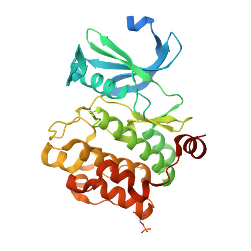
Proto-oncogene serine threonine kinase (PIM1) in complex with a consensus peptide and the JNK inhibitor V.

Filippakopoulos, P.Bullock, A.Fedorov, O.Pike, A.C.W.von Delft, F.Arrowsmith, C.H.Edwards, A.M.Bountra, C.Knapp, S.

To be published.

Macromolecule Content 

  • Total Structure Weight: 37.83 kDa 
  • Atom Count: 2,509 
  • Modeled Residue Count: 279 
  • Deposited Residue Count: 328 
  • Unique protein chains: 2

Macromolecules

Find similar proteins by:|  3D Structure
Entity ID: 1
MoleculeChains  Sequence LengthOrganismDetailsImage
Serine/threonine-protein kinase pim-1314Homo sapiensMutation(s): 1 
Gene Names: PIM1
EC: 2.7.11.1
UniProt & NIH Common Fund Data Resources
Find proteins for P11309 (Homo sapiens)
Explore P11309 
Go to UniProtKB:  P11309
PHAROS:  P11309
GTEx:  ENSG00000137193 
Entity Groups
Sequence Clusters30% Identity50% Identity70% Identity90% Identity95% Identity100% Identity
UniProt GroupP11309
Sequence Annotations
Expand
Reference Sequence
Find similar proteins by:  Sequence   |   3D Structure  
Entity ID: 2
MoleculeChains  Sequence LengthOrganismDetailsImage
Pimtide peptide14synthetic constructMutation(s): 0 

Small Molecules

Ligands 2 Unique
IDChains Name / Formula / InChI Key2D Diagram3D Interactions
JN5

Query on JN5


Download:Ideal Coordinates CCD File
D [auth A](2S)-1,3-benzothiazol-2-yl{2-[(2-pyridin-3-ylethyl)amino]pyrimidin-4-yl}ethanenitrile
C20 H16 N6 S
RCYPVQCPYKNSTG-OAHLLOKOSA-N
EDO

Query on EDO


Download:Ideal Coordinates CCD File
C [auth A]1,2-ETHANEDIOL
C2 H6 O2
LYCAIKOWRPUZTN-UHFFFAOYSA-N
Modified Residues  1 Unique
IDChains TypeFormula2D DiagramParent
SEP
Query on SEP
A
L-PEPTIDE LINKINGC3 H8 N O6 PSER
Binding Affinity Annotations 
IDSourceBinding Affinity
JN5 BindingDB:  3CY3 IC50: 80 (nM) from 1 assay(s)

Experimental Data & Validation

Experimental Data

  • Method: X-RAY DIFFRACTION
  • Resolution: 2.15 Å
  • R-Value Free:  0.205 (Depositor), 0.209 (DCC) 
  • R-Value Work:  0.163 (Depositor) 
  • R-Value Observed: 0.165 (Depositor) 
Space Group: P 65
Unit Cell:
Length ( Å )Angle ( ˚ )
a = 98.239α = 90
b = 98.239β = 90
c = 80.723γ = 120
Software Package:
Software NamePurpose
SCALAdata scaling
PHASERphasing
REFMACrefinement
PDB_EXTRACTdata extraction
CrystalCleardata collection
MOSFLMdata reduction

Structure Validation

View Full Validation Report


Ligand Structure Quality Assessment 


Entry History 

Revision History  (Full details and data files)

  • Version 1.0: 2008-07-15
    Type: Initial release
  • Version 1.1: 2011-07-13
    Changes: Advisory, Version format compliance
  • Version 1.2: 2017-10-25
    Changes: Author supporting evidence
  • Version 1.3: 2021-10-20
    Changes: Database references, Derived calculations
  • Version 1.4: 2022-02-16
    Changes: Database references, Derived calculations, Source and taxonomy, Structure summary
  • Version 1.5: 2023-08-30
    Changes: Data collection, Refinement description
  • Version 1.6: 2024-10-30
    Changes: Structure summary