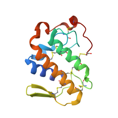
3CXI

Structure of BthTX-I complexed with alpha-tocopherol


Experimental Data Snapshot

  • Method: X-RAY DIFFRACTION
  • Resolution: 1.83 Å
  • R-Value Free: 0.243 
  • R-Value Work: 0.195 

wwPDB Validation   3D Report Full Report


Ligand Structure Quality Assessment 

Created with Raphaël 2.3.0Worse 01 BetterLigand structure goodness of fit to experimental dataBest fitted VITClick on this verticalbar to view details

This is version 1.2 of the entry. See complete history


Literature

Comparative structural studies on Lys49-phospholipases A(2) from Bothrops genus reveal their myotoxic site.

dos Santos, J.I.Soares, A.M.Fontes, M.R.

(2009) J Struct Biol 167: 106-116

  • DOI: https://doi.org/10.1016/j.jsb.2009.04.003
  • Primary Citation of Related Structures:  
    2Q2J, 3CXI, 3CYL

  • PubMed Abstract: 

    Phospholipases A(2) (PLA(2)s) are membrane-associated enzymes that hydrolyze phospholipids at the sn-2 position, releasing lysophospholipids and free fatty acids. Phospholipase A(2) homologues (Lys49-PLA(2)s) are highly myotoxic and cause extensive tissue damage despite not showing measurable catalytic activity. They are found in different snake venoms and represent one third of bothropic venom composition. The importance of these toxins during envenomation is related to the pronounced local myotoxic effect they induce since this effect is not neutralized by serum therapy. We present herein three structures of Lys49-PLA(2)s from Bothrops genus snake venom crystallized under the same conditions, two of which were grown in the presence of alpha-tocopherol (vitamin E). Comparative structural analysis of these and other Lys49-PLA(2)s showed two different patterns of oligomeric conformation that are related to the presence or absence of ligands in the hydrophobic channel. This work also confirms the biological dimer indicated by recent studies in which both C-termini are in the dimeric interface. In this configuration, we propose that the myotoxic site of these toxins is composed by the Lys 20, Lys115 and Arg118 residues. For the first time, a residue from the short-helix (Lys20) is suggested as a member of this site and the importance of Tyr119 residue to myotoxicity of bothropic Lys49-PLA(2)s is also discussed. These results support a complete hypothesis for these PLA(2)s myotoxic activity consistent with all findings on bothropic Lys49-PLA(2)s studied up to this moment, including crystallographic, bioinformatics, biochemical and biophysical data.


  • Organizational Affiliation

    Depto. de Física e Biofísica, Instituto de Biociências, UNESP, SP, Brazil.


Macromolecules
Find similar proteins by:  (by identity cutoff)  |  3D Structure
Entity ID: 1
MoleculeChains Sequence LengthOrganismDetailsImage
Myotoxic phospholipase A2-like
A, B
121Bothrops jararacussuMutation(s): 0 
UniProt
Find proteins for Q90249 (Bothrops jararacussu)
Explore Q90249 
Go to UniProtKB:  Q90249
Entity Groups  
Sequence Clusters30% Identity50% Identity70% Identity90% Identity95% Identity100% Identity
UniProt GroupQ90249
Sequence Annotations
Expand
  • Reference Sequence
Small Molecules
Ligands 3 Unique
IDChains Name / Formula / InChI Key2D Diagram3D Interactions
VIT
Query on VIT

Download Ideal Coordinates CCD File 
E [auth A],
I [auth B]
VITAMIN E
C29 H50 O2
GVJHHUAWPYXKBD-SYZUXVNWSA-N
PE4
Query on PE4

Download Ideal Coordinates CCD File 
H [auth B]2-{2-[2-(2-{2-[2-(2-ETHOXY-ETHOXY)-ETHOXY]-ETHOXY}-ETHOXY)-ETHOXY]-ETHOXY}-ETHANOL
C16 H34 O8
PJWQOENWHPEPKI-UHFFFAOYSA-N
SO4
Query on SO4

Download Ideal Coordinates CCD File 
C [auth A],
D [auth A],
F [auth B],
G [auth B]
SULFATE ION
O4 S
QAOWNCQODCNURD-UHFFFAOYSA-L
Experimental Data & Validation

Experimental Data

  • Method: X-RAY DIFFRACTION
  • Resolution: 1.83 Å
  • R-Value Free: 0.243 
  • R-Value Work: 0.195 
  • Space Group: P 1 21 1
Unit Cell:
Length ( Å )Angle ( ˚ )
a = 38.37α = 90
b = 70.13β = 102.23
c = 43.83γ = 90
Software Package:
Software NamePurpose
MAR345dtbdata collection
AMoREphasing
CNSrefinement
DENZOdata reduction
SCALEPACKdata scaling

Structure Validation

View Full Validation Report


Ligand Structure Quality Assessment 

Created with Raphaël 2.3.0Worse 01 BetterLigand structure goodness of fit to experimental dataBest fitted VITClick on this verticalbar to view details

Entry History 

Deposition Data

Revision History  (Full details and data files)

  • Version 1.0: 2009-05-05
    Type: Initial release
  • Version 1.1: 2011-07-13
    Changes: Version format compliance
  • Version 1.2: 2024-10-16
    Changes: Data collection, Database references, Derived calculations, Structure summary