3CWH | pdb_00003cwh

D-xylose Isomerase in complex with linear product, per-deuterated xylulose


Experimental Data Snapshot

  • Method: NEUTRON DIFFRACTION
  • Resolution: 2.20 Å
  • R-Value Free: 
    0.288 (Depositor) 
  • R-Value Observed: 
    0.237 (Depositor) 

Starting Model: experimental
View more details

wwPDB Validation 3D Report Full Report

Validation slider image for 3CWH

This is version 1.2 of the entry. See complete history

Literature

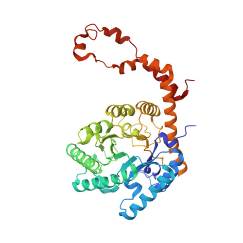
Hydrogen location in stages of an enzyme-catalyzed reaction: time-of-flight neutron structure of D-xylose isomerase with bound D-xylulose

Kovalevsky, A.Y.Katz, A.K.Carrell, H.L.Hanson, L.Mustyakimov, M.Fisher, S.Z.Coates, L.Schoenborn, B.P.Bunick, G.J.Glusker, J.P.Langan, P.

(2008) Biochemistry 47: 7595-7597

  • DOI: https://doi.org/10.1021/bi8005434
  • Primary Citation Related Structures: 
    3CWH

  • PubMed Abstract: 

    The time-of-flight neutron Laue technique has been used to determine the location of hydrogen atoms in the enzyme d-xylose isomerase (XI). The neutron structure of crystalline XI with bound product, d-xylulose, shows, unexpectedly, that O5 of d-xylulose is not protonated but is hydrogen-bonded to doubly protonated His54. Also, Lys289, which is neutral in native XI, is protonated (positively charged), while the catalytic water in native XI has become activated to a hydroxyl anion which is in the proximity of C1 and C2, the molecular site of isomerization of xylose. These findings impact our understanding of the reaction mechanism.


  • Organizational Affiliation
    • M888, Bioscience Division, Los Alamos National Laboratory, Los Alamos, New Mexico 87545, USA.

Macromolecule Content 

  • Total Structure Weight: 43.5 kDa 
  • Atom Count: 3,286 
  • Modeled Residue Count: 387 
  • Deposited Residue Count: 388 
  • Unique protein chains: 1

Macromolecules

Find similar proteins by:|  3D Structure
Entity ID: 1
MoleculeChains  Sequence LengthOrganismDetailsImage
Xylose isomerase388N/AMutation(s): 0 
EC: 5.3.1.5
UniProt
Find proteins for P24300 (Streptomyces pseudogriseolus)
Explore P24300 
Go to UniProtKB:  P24300
Entity Groups
Sequence Clusters30% Identity50% Identity70% Identity90% Identity95% Identity100% Identity
UniProt GroupP24300
Sequence Annotations
Expand
Reference Sequence

Experimental Data & Validation

Experimental Data

  • Method: NEUTRON DIFFRACTION
  • Resolution: 2.20 Å
  • R-Value Free:  0.288 (Depositor) 
  • R-Value Observed: 0.237 (Depositor) 
Space Group: I 2 2 2
Unit Cell:
Length ( Å )Angle ( ˚ )
a = 94.64α = 90
b = 99.97β = 90
c = 103.97γ = 90
Software Package:
Software NamePurpose
CNSrefinement
SHELXL-97refinement
d*TREKdata reduction
LAUENORMdata scaling
CNSphasing

Structure Validation

View Full Validation Report


Entry History 

Deposition Data

Revision History  (Full details and data files)

  • Version 1.0: 2008-08-05
    Type: Initial release
  • Version 1.1: 2011-07-13
    Changes: Non-polymer description, Version format compliance
  • Version 1.2: 2023-08-30
    Changes: Data collection, Database references, Derived calculations, Refinement description