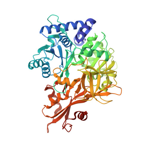
3CW9

4-Chlorobenzoyl-CoA Ligase/Synthetase in the Thioester-forming Conformation, bound to 4-chlorophenacyl-CoA


Experimental Data Snapshot

  • Method: X-RAY DIFFRACTION
  • Resolution: 2.00 Å
  • R-Value Free: 0.209 
  • R-Value Work: 0.166 
  • R-Value Observed: 0.168 

Starting Model: experimental
View more details

wwPDB Validation   3D Report Full Report


Ligand Structure Quality Assessment 


This is version 1.2 of the entry. See complete history


Literature

Structural characterization of a 140 degrees domain movement in the two-step reaction catalyzed by 4-chlorobenzoate:CoA ligase.

Reger, A.S.Wu, R.Dunaway-Mariano, D.Gulick, A.M.

(2008) Biochemistry 47: 8016-8025

  • DOI: https://doi.org/10.1021/bi800696y
  • Primary Citation of Related Structures:  
    3CW8, 3CW9

  • PubMed Abstract: 

    Members of the adenylate-forming family of enzymes play a role in the metabolism of halogenated aromatics and of short, medium, and long chain fatty acids, as well as in the biosynthesis of menaquinone, peptide antibiotics, and peptide siderophores. This family includes a subfamily of acyl- and aryl-CoA ligases that catalyze thioester synthesis through two half-reactions. A carboxylate substrate first reacts with ATP to form an acyl-adenylate. Subsequent to the release of the product PP i, the enzyme binds CoA, which attacks the activated acyl group to displace AMP. Structural and functional studies on different family members suggest that these enzymes alternate between two conformations during catalysis of the two half-reactions. Specifically, after the initial adenylation step, the C-terminal domain rotates by approximately 140 degrees to adopt a second conformation for thioester formation. Previously, we determined the structure of 4-chlorobenzoate:CoA ligase (CBL) in the adenylate forming conformation bound to 4-chlorobenzoate. We have determined two new crystal structures. We have determined the structure of CBL in the original adenylate-forming conformation, bound to the adenylate intermediate. Additionally, we have used a novel product analogue, 4-chlorophenacyl-CoA, to trap the enzyme in the thioester-forming conformation and determined this structure in a new crystal form. This work identifies a novel binding pocket for the CoA nucleotide. The structures presented herein provide the foundation for biochemical analyses presented in the accompanying manuscript in this issue [Wu et al. (2008) Biochemistry 47, 8026-8039]. The complete characterization of this enzyme allows us to provide an explanation for the use of the domain alternation strategy by these enzymes.


  • Organizational Affiliation

    Hauptman-Woodward Medical Research Institute, Buffalo, New York 14203, USA.


Macromolecules
Find similar proteins by:  (by identity cutoff)  |  3D Structure
Entity ID: 1
MoleculeChains Sequence LengthOrganismDetailsImage
4-chlorobenzoyl CoA ligase
A, B
504Alcaligenes sp.Mutation(s): 0 
EC: 6.2.1.33
UniProt
Find proteins for Q8GN86 (Alcaligenes sp. AL3007)
Explore Q8GN86 
Go to UniProtKB:  Q8GN86
Entity Groups  
Sequence Clusters30% Identity50% Identity70% Identity90% Identity95% Identity100% Identity
UniProt GroupQ8GN86
Sequence Annotations
Expand
  • Reference Sequence
Small Molecules
Ligands 5 Unique
IDChains Name / Formula / InChI Key2D Diagram3D Interactions
01A
Query on 01A

Download Ideal Coordinates CCD File 
F [auth A],
L [auth B]
4-Chlorophenacyl-coenzyme A
C29 H41 Cl N7 O17 P3 S
JKZHFXZXNBMAGG-VXAHOBLNSA-N
AMP
Query on AMP

Download Ideal Coordinates CCD File 
E [auth A],
K [auth B]
ADENOSINE MONOPHOSPHATE
C10 H14 N5 O7 P
UDMBCSSLTHHNCD-KQYNXXCUSA-N
EDO
Query on EDO

Download Ideal Coordinates CCD File 
G [auth A]1,2-ETHANEDIOL
C2 H6 O2
LYCAIKOWRPUZTN-UHFFFAOYSA-N
NO3
Query on NO3

Download Ideal Coordinates CCD File 
D [auth A],
I [auth B],
J [auth B]
NITRATE ION
N O3
NHNBFGGVMKEFGY-UHFFFAOYSA-N
MG
Query on MG

Download Ideal Coordinates CCD File 
C [auth A],
H [auth B]
MAGNESIUM ION
Mg
JLVVSXFLKOJNIY-UHFFFAOYSA-N
Experimental Data & Validation

Experimental Data

  • Method: X-RAY DIFFRACTION
  • Resolution: 2.00 Å
  • R-Value Free: 0.209 
  • R-Value Work: 0.166 
  • R-Value Observed: 0.168 
  • Space Group: P 21 21 21
Unit Cell:
Length ( Å )Angle ( ˚ )
a = 60.507α = 90
b = 87.789β = 90
c = 186.06γ = 90
Software Package:
Software NamePurpose
SSRLdata collection
MOLREPphasing
REFMACrefinement
MOSFLMdata reduction
SCALAdata scaling

Structure Validation

View Full Validation Report


Ligand Structure Quality Assessment 


Entry History 

Deposition Data

Revision History  (Full details and data files)

  • Version 1.0: 2008-09-02
    Type: Initial release
  • Version 1.1: 2011-07-13
    Changes: Advisory, Refinement description, Version format compliance
  • Version 1.2: 2023-08-30
    Changes: Data collection, Database references, Derived calculations, Refinement description