3CVE | pdb_00003cve

Crystal Structure of the carboxy terminus of Homer1


Experimental Data Snapshot

  • Method: X-RAY DIFFRACTION
  • Resolution: 1.75 Å
  • R-Value Free: 
    0.289 (Depositor), 0.338 (DCC) 
  • R-Value Work: 
    0.216 (Depositor), 0.286 (DCC) 
  • R-Value Observed: 
    0.220 (Depositor) 

wwPDB Validation 3D Report Full Report

Validation slider image for 3CVE

This is version 1.5 of the entry. See complete history

Literature

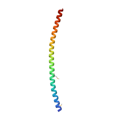
The postsynaptic density proteins Homer and Shank form a polymeric network structure.

Hayashi, M.K.Tang, C.Verpelli, C.Narayanan, R.Stearns, M.H.Xu, R.M.Li, H.Sala, C.Hayashi, Y.

(2009) Cell 137: 159-171

  • DOI: https://doi.org/10.1016/j.cell.2009.01.050
  • Primary Citation Related Structures: 
    3CVE, 3CVF

  • PubMed Abstract: 

    The postsynaptic density (PSD) is crucial for synaptic functions, but the molecular architecture retaining its structure and components remains elusive. Homer and Shank are among the most abundant scaffolding proteins in the PSD, working synergistically for maturation of dendritic spines. Here, we demonstrate that Homer and Shank, together, form a mesh-like matrix structure. Crystallographic analysis of this region revealed a pair of parallel dimeric coiled coils intercalated in a tail-to-tail fashion to form a tetramer, giving rise to the unique configuration of a pair of N-terminal EVH1 domains at each end of the coiled coil. In neurons, the tetramerization is required for structural integrity of the dendritic spines and recruitment of proteins to synapses. We propose that the Homer-Shank complex serves as a structural framework and as an assembly platform for other PSD proteins.


  • Organizational Affiliation
    • RIKEN-MIT Neuroscience Research Center, The Picower Institute for Learning and Memory, Department of Brain and Cognitive Sciences, Massachusetts Institute of Technology, Cambridge, MA 02139, USA. hayashim@mit.edu

Macromolecule Content 

  • Total Structure Weight: 33.86 kDa 
  • Atom Count: 2,417 
  • Modeled Residue Count: 261 
  • Deposited Residue Count: 288 
  • Unique protein chains: 1

Macromolecules

Find similar proteins by:|  3D Structure
Entity ID: 1
MoleculeChains  Sequence LengthOrganismDetailsImage
Homer protein homolog 1
A, B, C, D
72Rattus norvegicusMutation(s): 1 
Gene Names: Homer1HomerVesl
UniProt
Find proteins for Q9Z214 (Rattus norvegicus)
Explore Q9Z214 
Go to UniProtKB:  Q9Z214
Entity Groups
Sequence Clusters30% Identity50% Identity70% Identity90% Identity95% Identity100% Identity
UniProt GroupQ9Z214
Sequence Annotations
Expand
Reference Sequence

Small Molecules

Modified Residues  1 Unique
IDChains TypeFormula2D DiagramParent
MSE
Query on MSE
A, B, C, D
L-PEPTIDE LINKINGC5 H11 N O2 SeMET

Experimental Data & Validation

Experimental Data

  • Method: X-RAY DIFFRACTION
  • Resolution: 1.75 Å
  • R-Value Free:  0.289 (Depositor), 0.338 (DCC) 
  • R-Value Work:  0.216 (Depositor), 0.286 (DCC) 
  • R-Value Observed: 0.220 (Depositor) 
Space Group: P 1 21 1
Unit Cell:
Length ( Å )Angle ( ˚ )
a = 39.014α = 90
b = 62.508β = 102.14
c = 57.526γ = 90
Software Package:
Software NamePurpose
DENZOdata reduction
SCALEPACKdata scaling
SHARPphasing
REFMACrefinement
PDB_EXTRACTdata extraction

Structure Validation

View Full Validation Report


Entry History 

Deposition Data

Revision History  (Full details and data files)

  • Version 1.0: 2009-03-31
    Type: Initial release
  • Version 1.1: 2011-07-13
    Changes: Version format compliance
  • Version 1.2: 2017-06-07
    Changes: Refinement description
  • Version 1.3: 2017-10-25
    Changes: Refinement description
  • Version 1.4: 2021-10-20
    Changes: Database references, Derived calculations
  • Version 1.5: 2024-10-16
    Changes: Data collection, Structure summary