3CV7 | pdb_00003cv7

Crystal structure of porcine aldehyde reductase ternary complex


Experimental Data Snapshot

  • Method: X-RAY DIFFRACTION
  • Resolution: 2.41 Å
  • R-Value Free: 
    0.247 (Depositor), 0.245 (DCC) 
  • R-Value Work: 
    0.191 (Depositor), 0.192 (DCC) 
  • R-Value Observed: 
    0.194 (Depositor) 

Starting Model: experimental
View more details

wwPDB Validation 3D Report Full Report

Validation slider image for 3CV7

Ligand Structure Quality Assessment 


This is version 1.2 of the entry. See complete history

Literature

Structure of aldehyde reductase in ternary complex with coenzyme and the potent 20alpha-hydroxysteroid dehydrogenase inhibitor 3,5-dichlorosalicylic acid: Implications for inhibitor binding and selectivity

Carbone, V.Chung, R.Endo, S.Hara, A.El-Kabbani, O.

(2008) Arch Biochem Biophys 479: 82-87

  • DOI: https://doi.org/10.1016/j.abb.2008.08.014
  • Primary Citation Related Structures: 
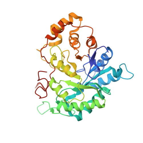
    3CV7

  • PubMed Abstract: 

    The structure of aldehyde reductase (ALR1) in ternary complex with the coenzyme NADPH and 3,5-dichlorosalicylic acid (DCL), a potent inhibitor of human 20alpha-hydroxysteroid dehydrogenase (AKR1C1), was determined at a resolution of 2.41A. The inhibitor formed a network of hydrogen bonds with the active site residues Trp22, Tyr50, His113, Trp114 and Arg312. Molecular modelling calculations together with inhibitory activity measurements indicated that DCL was a less potent inhibitor of ALR1 (256-fold) when compared to AKR1C1. In AKR1C1, the inhibitor formed a 10-fold stronger binding interaction with the catalytic residue (Tyr55), non-conserved hydrogen bonding interaction with His222, and additional van der Waals contacts with the non-conserved C-terminal residues Leu306, Leu308 and Phe311 that contribute to the inhibitor's selectivity advantage for AKR1C1 over ALR1.


  • Organizational Affiliation
    • Medicinal Chemistry and Drug Action, Monash Institute of Pharmaceutical Sciences, Parkville, Victoria, Australia.

Macromolecule Content 

  • Total Structure Weight: 37.96 kDa 
  • Atom Count: 2,899 
  • Modeled Residue Count: 322 
  • Deposited Residue Count: 325 
  • Unique protein chains: 1

Macromolecules

Find similar proteins by:|  3D Structure
Entity ID: 1
MoleculeChains  Sequence LengthOrganismDetailsImage
Alcohol dehydrogenase325Sus scrofaMutation(s): 0 
EC: 1.1.1.2 (PDB Primary Data), 1.6 (UniProt), 1.1.1.20 (UniProt), 1.1.1.19 (UniProt), 1.1.1.372 (UniProt), 1.1.1.54 (UniProt)
UniProt
Find proteins for P50578 (Sus scrofa)
Explore P50578 
Go to UniProtKB:  P50578
Entity Groups
Sequence Clusters30% Identity50% Identity70% Identity90% Identity95% Identity100% Identity
UniProt GroupP50578
Sequence Annotations
Expand
Reference Sequence

Small Molecules

Binding Affinity Annotations 
IDSourceBinding Affinity
C2U BindingDB:  3CV7 IC50: 1.13e+4 (nM) from 1 assay(s)

Experimental Data & Validation

Experimental Data

  • Method: X-RAY DIFFRACTION
  • Resolution: 2.41 Å
  • R-Value Free:  0.247 (Depositor), 0.245 (DCC) 
  • R-Value Work:  0.191 (Depositor), 0.192 (DCC) 
  • R-Value Observed: 0.194 (Depositor) 
Space Group: P 65 2 2
Unit Cell:
Length ( Å )Angle ( ˚ )
a = 67.385α = 90
b = 67.385β = 90
c = 246.106γ = 120
Software Package:
Software NamePurpose
REFMACrefinement
MAR345dtbdata collection
AUTOMARdata reduction
MOLREPphasing

Structure Validation

View Full Validation Report


Ligand Structure Quality Assessment 


Entry History 

Deposition Data

Revision History  (Full details and data files)

  • Version 1.0: 2008-10-28
    Type: Initial release
  • Version 1.1: 2011-07-13
    Changes: Version format compliance
  • Version 1.2: 2023-11-01
    Changes: Data collection, Database references, Derived calculations, Refinement description