3CRV | pdb_00003crv

XPD_Helicase


Experimental Data Snapshot

  • Method: X-RAY DIFFRACTION
  • Resolution: 2.00 Å
  • R-Value Free: 
    0.260 (Depositor), 0.254 (DCC) 
  • R-Value Work: 
    0.222 (Depositor), 0.214 (DCC) 
  • R-Value Observed: 
    0.227 (Depositor) 

wwPDB Validation 3D Report Full Report

Validation slider image for 3CRV

This is version 1.5 of the entry. See complete history

Literature

XPD helicase structures and activities: insights into the cancer and aging phenotypes from XPD mutations.

Fan, L.Fuss, J.O.Cheng, Q.J.Arvai, A.S.Hammel, M.Roberts, V.A.Cooper, P.K.Tainer, J.A.

(2008) Cell 133: 789-800

  • DOI: https://doi.org/10.1016/j.cell.2008.04.030
  • Primary Citation Related Structures: 
    3CRV, 3CRW

  • PubMed Abstract: 

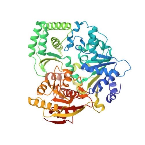
    Mutations in XPD helicase, required for nucleotide excision repair (NER) as part of the transcription/repair complex TFIIH, cause three distinct phenotypes: cancer-prone xeroderma pigmentosum (XP), or aging disorders Cockayne syndrome (CS), and trichothiodystrophy (TTD). To clarify molecular differences underlying these diseases, we determined crystal structures of the XPD catalytic core from Sulfolobus acidocaldarius and measured mutant enzyme activities. Substrate-binding grooves separate adjacent Rad51/RecA-like helicase domains (HD1, HD2) and an arch formed by 4FeS and Arch domains. XP mutations map along the HD1 ATP-binding edge and HD2 DNA-binding channel and impair helicase activity essential for NER. XP/CS mutations both impair helicase activity and likely affect HD2 functional movement. TTD mutants lose or retain helicase activity but map to sites in all four domains expected to cause framework defects impacting TFIIH integrity. These results provide a foundation for understanding disease consequences of mutations in XPD and related 4Fe-4S helicases including FancJ.


  • Organizational Affiliation
    • Department of Molecular Biology, Skaggs Institute of Chemical Biology, The Scripps Research Institute, La Jolla, CA 92037, USA.

Macromolecule Content 

  • Total Structure Weight: 65.14 kDa 
  • Atom Count: 4,819 
  • Modeled Residue Count: 551 
  • Deposited Residue Count: 551 
  • Unique protein chains: 1

Macromolecules

Find similar proteins by:|  3D Structure
Entity ID: 1
MoleculeChains  Sequence LengthOrganismDetailsImage
XPD/Rad3 related DNA helicase551Sulfolobus acidocaldariusMutation(s): 1 
Gene Names: Saci_0192
EC: 3 (PDB Primary Data), 5.6.2.3 (UniProt)
UniProt
Find proteins for Q4JC68 (Sulfolobus acidocaldarius (strain ATCC 33909 / DSM 639 / JCM 8929 / NBRC 15157 / NCIMB 11770))
Explore Q4JC68 
Go to UniProtKB:  Q4JC68
Entity Groups
Sequence Clusters30% Identity50% Identity70% Identity90% Identity95% Identity100% Identity
UniProt GroupQ4JC68
Sequence Annotations
Expand
Reference Sequence

Small Molecules

Ligands 4 Unique
IDChains Name / Formula / InChI Key2D Diagram3D Interactions
SF4

Query on SF4


Download:Ideal Coordinates CCD File
C [auth A]IRON/SULFUR CLUSTER
Fe4 S4
LJBDFODJNLIPKO-UHFFFAOYSA-N
FLC

Query on FLC


Download:Ideal Coordinates CCD File
B [auth A]CITRATE ANION
C6 H5 O7
KRKNYBCHXYNGOX-UHFFFAOYSA-K
GOL

Query on GOL


Download:Ideal Coordinates CCD File
K [auth A]GLYCEROL
C3 H8 O3
PEDCQBHIVMGVHV-UHFFFAOYSA-N
IPA

Query on IPA


Download:Ideal Coordinates CCD File
D [auth A]
E [auth A]
F [auth A]
G [auth A]
H [auth A]
D [auth A],
E [auth A],
F [auth A],
G [auth A],
H [auth A],
I [auth A],
J [auth A]
ISOPROPYL ALCOHOL
C3 H8 O
KFZMGEQAYNKOFK-UHFFFAOYSA-N

Experimental Data & Validation

Experimental Data

  • Method: X-RAY DIFFRACTION
  • Resolution: 2.00 Å
  • R-Value Free:  0.260 (Depositor), 0.254 (DCC) 
  • R-Value Work:  0.222 (Depositor), 0.214 (DCC) 
  • R-Value Observed: 0.227 (Depositor) 
Space Group: P 21 21 21
Unit Cell:
Length ( Å )Angle ( ˚ )
a = 53.542α = 90
b = 70.222β = 90
c = 144.293γ = 90
Software Package:
Software NamePurpose
CNSrefinement
HKL-2000data collection
HKL-2000data reduction
HKL-2000data scaling
SOLVEphasing

Structure Validation

View Full Validation Report


Entry History 

Deposition Data

Revision History  (Full details and data files)

  • Version 1.0: 2008-06-10
    Type: Initial release
  • Version 1.1: 2011-07-13
    Changes: Non-polymer description, Version format compliance
  • Version 1.2: 2018-01-24
    Changes: Structure summary
  • Version 1.3: 2018-09-05
    Changes: Data collection, Source and taxonomy, Structure summary
  • Version 1.4: 2021-10-20
    Changes: Database references, Derived calculations
  • Version 1.5: 2024-02-21
    Changes: Data collection