3CMY

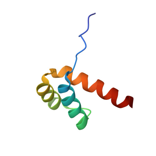
Structure of a homeodomain in complex with DNA


Experimental Data Snapshot

  • Method: X-RAY DIFFRACTION
  • Resolution: 1.95 Å
  • R-Value Free: 0.213 
  • R-Value Work: 0.172 
  • R-Value Observed: 0.174 

Starting Model: experimental
View more details

wwPDB Validation   3D Report Full Report


This is version 1.3 of the entry. See complete history


Literature

Structural Basis for DNA Recognition by the Human PAX3 Homeodomain.

Birrane, G.Soni, A.Ladias, J.A.

(2009) Biochemistry 48: 1148-1155

  • DOI: https://doi.org/10.1021/bi802052y
  • Primary Citation of Related Structures:  
    3CMY

  • PubMed Abstract: 

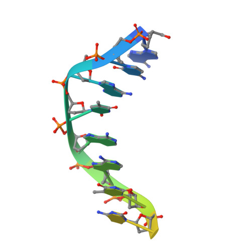
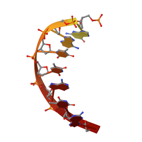
    The transcription regulatory protein PAX3 binds to cognate DNA sequences through two DNA-binding domains, a paired domain and a homeodomain, and has important functions during neurogenesis and myogenesis. In humans, mutations in the PAX3 gene cause Waardenburg syndrome, whereas a chromosomal translocation that generates a PAX3-FOXO1 fusion gene is associated with the development of alveolar rhabdomyosarcoma. We have determined the crystal structure of the human PAX3 homeodomain in complex with a palindromic DNA containing two inverted TAATC sequences at 1.95 A resolution. Two homeodomains bind to DNA as a symmetric dimer, inducing a 3 degrees bend in the DNA helix. The N-terminal arm of the homeodomain inserts into the minor groove and makes direct and water-mediated interactions with bases and the sugar-phosphate backbone. The recognition helix fits directly into the major groove, and an elaborate network of structurally conserved water molecules mediates the majority of protein-DNA interactions. The structure elucidates the role of serine 50 in selection of the CG sequence immediately 3' of the TAAT motif by PAX class homeodomains and provides insights into the molecular mechanisms by which certain Waardenburg syndrome-associated missense mutations could destabilize the fold of the PAX3 homeodomain whereas others could affect its interaction with DNA.


  • Organizational Affiliation

    Molecular Medicine Laboratory and Macromolecular Crystallography Unit, Division of Experimental Medicine, Harvard Medical School, Boston, Massachusetts 02115, USA.


Macromolecules

Find similar proteins by:  (by identity cutoff)  |  3D Structure
Entity ID: 1
MoleculeChains Sequence LengthOrganismDetailsImage
Paired box protein Pax-361Homo sapiensMutation(s): 0 
Gene Names: PAX3
UniProt & NIH Common Fund Data Resources
Find proteins for P23760 (Homo sapiens)
Explore P23760 
Go to UniProtKB:  P23760
PHAROS:  P23760
GTEx:  ENSG00000135903 
Entity Groups  
Sequence Clusters30% Identity50% Identity70% Identity90% Identity95% Identity100% Identity
UniProt GroupP23760
Sequence Annotations
Expand
  • Reference Sequence

Find similar nucleic acids by:  Sequence   |   3D Structure  

Entity ID: 2
MoleculeChains LengthOrganismImage
5'-D(*DAP*DCP*DAP*DTP*DAP*DAP*DP*DCP*DGP*DAP*DTP*DTP*DAP*DC)-3'14N/A
Sequence Annotations
Expand
  • Reference Sequence

Find similar nucleic acids by:  Sequence   |   3D Structure  

Entity ID: 3
MoleculeChains LengthOrganismImage
5'-D(*DTP*DGP*DTP*DAP*DAP*DTP*DCP*DGP*DAP*DTP*DTP*DAP*DTP*DG)-3'14N/A
Sequence Annotations
Expand
  • Reference Sequence
Small Molecules
Ligands 1 Unique
IDChains Name / Formula / InChI Key2D Diagram3D Interactions
EDO
Query on EDO

Download Ideal Coordinates CCD File 
D [auth A]1,2-ETHANEDIOL
C2 H6 O2
LYCAIKOWRPUZTN-UHFFFAOYSA-N
Experimental Data & Validation

Experimental Data

  • Method: X-RAY DIFFRACTION
  • Resolution: 1.95 Å
  • R-Value Free: 0.213 
  • R-Value Work: 0.172 
  • R-Value Observed: 0.174 
  • Space Group: C 2 2 21
Unit Cell:
Length ( Å )Angle ( ˚ )
a = 43.325α = 90
b = 62.243β = 90
c = 92.556γ = 90
Software Package:
Software NamePurpose
REFMACrefinement
HKL-2000data collection
HKL-2000data reduction
HKL-2000data scaling
MOLREPphasing

Structure Validation

View Full Validation Report


Entry History 

Deposition Data

Revision History  (Full details and data files)

  • Version 1.0: 2009-02-17
    Type: Initial release
  • Version 1.1: 2011-07-13
    Changes: Advisory, Refinement description, Version format compliance
  • Version 1.2: 2024-02-21
    Changes: Data collection, Database references, Derived calculations
  • Version 1.3: 2024-04-03
    Changes: Refinement description