3CM3 | pdb_00003cm3

High Resolution Crystal Structure of the Vaccinia Virus Dual-Specificity Phosphatase VH1


Experimental Data Snapshot

  • Method: X-RAY DIFFRACTION
  • Resolution: 1.32 Å
  • R-Value Free: 
    0.185 (Depositor), 0.179 (DCC) 
  • R-Value Work: 
    0.171 (Depositor), 0.167 (DCC) 
  • R-Value Observed: 
    0.172 (Depositor) 

Starting Model: experimental
View more details

wwPDB Validation 3D Report Full Report

Validation slider image for 3CM3

This is version 1.3 of the entry. See complete history

Literature

Dimeric Quaternary Structure of the Prototypical Dual Specificity Phosphatase VH1.

Koksal, A.C.Nardozzi, J.D.Cingolani, G.

(2009) J Biological Chem 284: 10129-10137

  • DOI: https://doi.org/10.1074/jbc.M808362200
  • Primary Citation Related Structures: 
    3CM3

  • PubMed Abstract: 

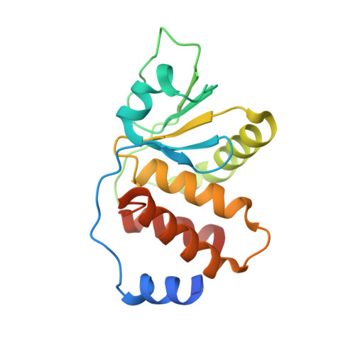
    The Vaccinia virus H1 gene product, VH1, is a dual specificity phosphatase that down-regulates the cellular antiviral response by dephosphorylating STAT1. The crystal structure of VH1, determined at 1.32 A resolution, reveals a novel dimeric quaternary structure, which exposes two active sites spaced approximately 39 A away from each other. VH1 forms a stable dimer via an extensive domain swap of the N-terminal helix (residues 1-20). In vitro, VH1 can dephosphorylate activated STAT1, in a reaction that is competed by the nuclear transport adapter importin alpha5. Interestingly, VH1 is inactive with respect to STAT1 bound to DNA, suggesting that the viral phosphatase acts predominantly on the cytoplasmic pool of activated STAT1. We propose that the dimeric quaternary structure of VH1 is essential for specific recognition of activated STAT1, which prevents its nuclear translocation, thus blocking interferon-gamma signal transduction and antiviral response.


  • Organizational Affiliation
    • Department of Biochemistry and Molecular Biology, SUNY Upstate Medical University, Syracuse, New York 13210, USA.

Macromolecule Content 

  • Total Structure Weight: 20.51 kDa 
  • Atom Count: 1,679 
  • Modeled Residue Count: 164 
  • Deposited Residue Count: 176 
  • Unique protein chains: 1

Macromolecules

Find similar proteins by:|  3D Structure
Entity ID: 1
MoleculeChains  Sequence LengthOrganismDetailsImage
Dual specificity protein phosphatase176N/AMutation(s): 1 
Gene Names: H1 ORFOPG106
EC: 3.1.3.48 (PDB Primary Data), 3.1.3 (PDB Primary Data)
UniProt
Find proteins for P07239 (Vaccinia virus (strain Western Reserve))
Explore P07239 
Go to UniProtKB:  P07239
Entity Groups
Sequence Clusters30% Identity50% Identity70% Identity90% Identity95% Identity100% Identity
UniProt GroupP07239
Sequence Annotations
Expand
Reference Sequence

Experimental Data & Validation

Experimental Data

  • Method: X-RAY DIFFRACTION
  • Resolution: 1.32 Å
  • R-Value Free:  0.185 (Depositor), 0.179 (DCC) 
  • R-Value Work:  0.171 (Depositor), 0.167 (DCC) 
  • R-Value Observed: 0.172 (Depositor) 
Space Group: C 2 2 21
Unit Cell:
Length ( Å )Angle ( ˚ )
a = 63.816α = 90
b = 38.69β = 90
c = 134.987γ = 90
Software Package:
Software NamePurpose
REFMACrefinement
ADSCdata collection
HKL-2000data reduction
HKL-2000data scaling
MOLREPphasing

Structure Validation

View Full Validation Report


Entry History 

Deposition Data

Revision History  (Full details and data files)

  • Version 1.0: 2009-02-10
    Type: Initial release
  • Version 1.1: 2011-07-13
    Changes: Version format compliance
  • Version 1.2: 2021-10-20
    Changes: Database references, Derived calculations
  • Version 1.3: 2023-08-30
    Changes: Data collection, Refinement description