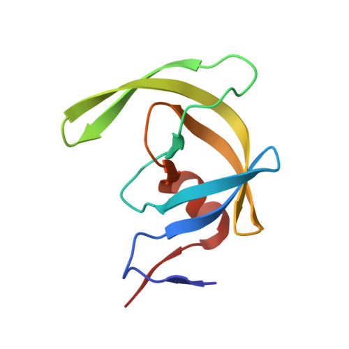
3CKT

HIV-1 protease in complex with a dimethylallyl decorated pyrrolidine based inhibitor (orthorombic space group)


Experimental Data Snapshot

  • Method: X-RAY DIFFRACTION
  • Resolution: 1.65 Å
  • R-Value Free: 0.239 
  • R-Value Work: 0.183 
  • R-Value Observed: 0.185 

Starting Model: experimental
View more details

wwPDB Validation   3D Report Full Report


Ligand Structure Quality Assessment 


This is version 1.3 of the entry. See complete history


Literature

Two Solutions for the Same Problem: Multiple Binding Modes of Pyrrolidine-Based HIV-1 Protease Inhibitors

Blum, A.Bottcher, J.Dorr, S.Heine, A.Klebe, G.Diederich, W.E.

(2011) J Mol Biol 410: 745-755

  • DOI: https://doi.org/10.1016/j.jmb.2011.04.052
  • Primary Citation of Related Structures:  
    2ZGA, 3CKT

  • PubMed Abstract: 

    Structure-based drug design is an integral part of industrial and academic drug discovery projects. Initial lead structures are, in general, optimized in terms of affinity using iterative cycles comprising synthesis, biological evaluation, computational methods, and structural analysis. X-ray crystallography commonly suggests the existence of a single well-defined state, termed binding mode, which is generally assumed to be consistent in a series of similar ligands and therefore used for the following optimization process. During the further development of symmetrically disubstituted 3,4-amino-pyrrolidines as human immunodeficiency virus type 1 protease inhibitors, we discovered that, by modification of the P1/P1' moieties of our lead structure, the activity of the inhibitors towards the active-site mutation Ile84Val was altered, however, not being explainable with the initial underlying structure-activity relationship. The cocrystallization of the most potent derivative in complex with the human immunodeficiency virus type 1 protease surprisingly led to two different crystal forms (P2(1)2(1)2(1) and P6(1)22). Structural analysis revealed two completely different binding modes; the interaction of the pyrrolidine nitrogen atom with the catalytic aspartates remains as the only similarity. The study presented clearly demonstrates that structural biology has to escort the entire lead optimization process not to fail by an initially observed binding orientation.


  • Organizational Affiliation

    Institut für Pharmazeutische Chemie, Philipps-Universität Marburg, Marburg, Germany.


Macromolecules
Find similar proteins by:  (by identity cutoff)  |  3D Structure
Entity ID: 1
MoleculeChains Sequence LengthOrganismDetailsImage
Protease
A, B
99Human immunodeficiency virus 1Mutation(s): 0 
Gene Names: gag-pol
EC: 3.4.23.16
UniProt
Find proteins for P03367 (Human immunodeficiency virus type 1 group M subtype B (isolate BRU/LAI))
Explore P03367 
Go to UniProtKB:  P03367
Entity Groups  
Sequence Clusters30% Identity50% Identity70% Identity90% Identity95% Identity100% Identity
UniProt GroupP03367
Sequence Annotations
Expand
  • Reference Sequence
Small Molecules
Binding Affinity Annotations 
IDSourceBinding Affinity
YDP PDBBind:  3CKT Ki: 480 (nM) from 1 assay(s)
Experimental Data & Validation

Experimental Data

  • Method: X-RAY DIFFRACTION
  • Resolution: 1.65 Å
  • R-Value Free: 0.239 
  • R-Value Work: 0.183 
  • R-Value Observed: 0.185 
  • Space Group: P 21 21 21
Unit Cell:
Length ( Å )Angle ( ˚ )
a = 52.018α = 90
b = 57.581β = 90
c = 61.365γ = 90
Software Package:
Software NamePurpose
SHELXmodel building
SHELXL-97refinement
MAR345dtbdata collection
HKL-2000data reduction
HKL-2000data scaling
PHASERphasing

Structure Validation

View Full Validation Report


Ligand Structure Quality Assessment 


Entry History 

Deposition Data

Revision History  (Full details and data files)

  • Version 1.0: 2009-03-24
    Type: Initial release
  • Version 1.1: 2011-07-13
    Changes: Version format compliance
  • Version 1.2: 2011-08-10
    Changes: Database references, Structure summary
  • Version 1.3: 2023-11-01
    Changes: Data collection, Database references, Derived calculations, Refinement description