3CJB | pdb_00003cjb

Actin dimer cross-linked by V. cholerae MARTX toxin and complexed with Gelsolin-segment 1


Experimental Data Snapshot

  • Method: X-RAY DIFFRACTION
  • Resolution: 3.21 Å
  • R-Value Free: 
    0.269 (Depositor), 0.280 (DCC) 
  • R-Value Work: 
    0.245 (Depositor), 0.260 (DCC) 
  • R-Value Observed: 
    0.246 (Depositor) 

wwPDB Validation   3D Report Full Report


Ligand Structure Quality Assessment 

Created with Raphaël 2.3.0Worse 01 BetterLigand structure goodness of fit to experimental dataBest fitted ATPClick on this verticalbar to view details

This is version 1.3 of the entry. See complete history


Literature

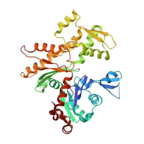
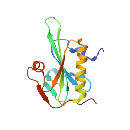
Connecting actin monomers by iso-peptide bond is a toxicity mechanism of the Vibrio cholerae MARTX toxin.

Kudryashov, D.S.Durer, Z.A.Ytterberg, A.J.Sawaya, M.R.Pashkov, I.Prochazkova, K.Yeates, T.O.Loo, R.R.Loo, J.A.Satchell, K.J.Reisler, E.

(2008) Proc Natl Acad Sci U S A 105: 18537-18542

  • DOI: https://doi.org/10.1073/pnas.0808082105
  • Primary Citation of Related Structures:  
    3CJB, 3CJC

  • PubMed Abstract: 

    The Gram-negative bacterium Vibrio cholerae is the causative agent of a severe diarrheal disease that afflicts three to five million persons annually, causing up to 200,000 deaths. Nearly all V. cholerae strains produce a large multifunctional-autoprocessing RTX toxin (MARTX(Vc)), which contributes significantly to the pathogenesis of cholera in model systems. The actin cross-linking domain (ACD) of MARTX(Vc) directly catalyzes a covalent cross-linking of monomeric G-actin into oligomeric chains and causes cell rounding, but the nature of the cross-linked bond and the mechanism of the actin cytoskeleton disruption remained elusive. To elucidate the mechanism of ACD action and effect on actin, we identified the covalent cross-link bond between actin protomers using limited proteolysis, X-ray crystallography, and mass spectrometry. We report here that ACD catalyzes the formation of an intermolecular iso-peptide bond between residues E270 and K50 located in the hydrophobic and the DNaseI-binding loops of actin, respectively. Mutagenesis studies confirm that no other residues on actin can be cross-linked by ACD both in vitro and in vivo. This cross-linking locks actin protomers into an orientation different from that of F-actin, resulting in strong inhibition of actin polymerization. This report describes a microbial toxin mechanism acting via iso-peptide bond cross-linking between host proteins and is, to the best of our knowledge, the only known example of a peptide linkage between nonterminal glutamate and lysine side chains.


  • Organizational Affiliation

    Department of Chemistry and Biochemistry and Molecular Biology Institute, University of California, Los Angeles, CA 90095, USA.


Macromolecules
Find similar proteins by:  (by identity cutoff)  |  3D Structure
Entity ID: 1
MoleculeChains Sequence LengthOrganismDetailsImage
Actin, alpha skeletal muscle377Oryctolagus cuniculusMutation(s): 0 
EC: 3.6.4
UniProt
Find proteins for P68135 (Oryctolagus cuniculus)
Explore P68135 
Go to UniProtKB:  P68135
Entity Groups  
Sequence Clusters30% Identity50% Identity70% Identity90% Identity95% Identity100% Identity
UniProt GroupP68135
Sequence Annotations
Expand
  • Reference Sequence
Find similar proteins by:  (by identity cutoff)  |  3D Structure
Entity ID: 2
MoleculeChains Sequence LengthOrganismDetailsImage
GelsolinB [auth G]125Homo sapiensMutation(s): 0 
Gene Names: GSN
UniProt & NIH Common Fund Data Resources
Find proteins for P06396 (Homo sapiens)
Explore P06396 
Go to UniProtKB:  P06396
PHAROS:  P06396
GTEx:  ENSG00000148180 
Entity Groups  
Sequence Clusters30% Identity50% Identity70% Identity90% Identity95% Identity100% Identity
UniProt GroupP06396
Sequence Annotations
Expand
  • Reference Sequence
Experimental Data & Validation

Experimental Data

  • Method: X-RAY DIFFRACTION
  • Resolution: 3.21 Å
  • R-Value Free:  0.269 (Depositor), 0.280 (DCC) 
  • R-Value Work:  0.245 (Depositor), 0.260 (DCC) 
  • R-Value Observed: 0.246 (Depositor) 
Space Group: P 21 21 21
Unit Cell:
Length ( Å )Angle ( ˚ )
a = 56.696α = 90
b = 69.54β = 90
c = 182.106γ = 90
Software Package:
Software NamePurpose
DENZOdata reduction
SCALEPACKdata scaling
PHASERphasing
REFMACrefinement
PDB_EXTRACTdata extraction
BOSdata collection

Structure Validation

View Full Validation Report


Ligand Structure Quality Assessment 

Created with Raphaël 2.3.0Worse 01 BetterLigand structure goodness of fit to experimental dataBest fitted ATPClick on this verticalbar to view details

Entry History 

Deposition Data

Revision History  (Full details and data files)

  • Version 1.0: 2008-03-25
    Type: Initial release
  • Version 1.1: 2011-07-13
    Changes: Advisory, Version format compliance
  • Version 1.2: 2017-10-25
    Changes: Refinement description
  • Version 1.3: 2024-02-21
    Changes: Data collection, Database references, Derived calculations