3CFC | pdb_00003cfc

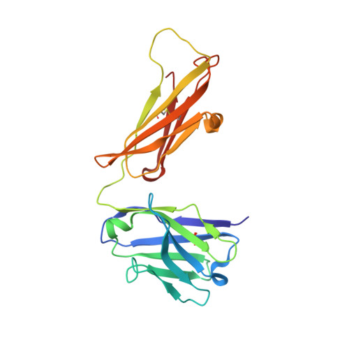
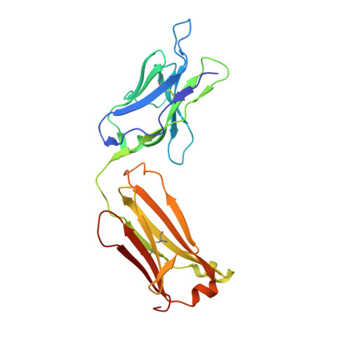
High-resolution structure of blue fluorescent antibody EP2-19G2


Experimental Data Snapshot

  • Method: X-RAY DIFFRACTION
  • Resolution: 1.70 Å
  • R-Value Free: 
    0.231 (Depositor), 0.294 (DCC) 
  • R-Value Work: 
    0.191 (Depositor), 0.261 (DCC) 
  • R-Value Observed: 
    0.193 (Depositor) 

Starting Model: experimental
View more details

wwPDB Validation 3D Report Full Report

Validation slider image for 3CFC

This is version 1.3 of the entry. See complete history. 

Literature

Deeply inverted electron-hole recombination in a luminescent antibody-stilbene complex.

Debler, E.W., Kaufmann, G.F., Meijler, M.M., Heine, A., Mee, J.M., Pljevaljcic, G., Di Bilio, A.J., Schultz, P.G., Millar, D.P., Janda, K.D., Wilson, I.A., Gray, H.B., Lerner, R.A.

(2008) Science 319: 1232-1235

  • DOI: https://doi.org/10.1126/science.1153445
  • Primary Citation Related Structures: 
    3CFB, 3CFC, 3CFD, 3CFE

  • PubMed Abstract: 

    The blue-emissive antibody EP2-19G2 that has been elicited against trans-stilbene has unprecedented ability to produce bright luminescence and has been used as a biosensor in various applications. We show that the prolonged luminescence is not stilbene fluorescence. Instead, the emissive species is a charge-transfer excited complex of an anionic stilbene and a cationic, parallel pi-stacked tryptophan. Upon charge recombination, this complex generates exceptionally bright blue light. Complex formation is enabled by a deeply penetrating ligand-binding pocket, which in turn results from a noncanonical interface between the two variable domains of the antibody.


  • Organizational Affiliation: 
    • Department of Molecular Biology, Scripps Research Institute, La Jolla, CA 92037, USA.

Macromolecule Content 

  • Total Structure Weight: 46.92 kDa 
  • Atom Count: 3,703 
  • Modeled Residue Count: 432 
  • Deposited Residue Count: 432 
  • Unique protein chains: 2

Macromolecules

Find similar proteins by:|  3D Structure
Entity ID: 1
MoleculeChains  Sequence LengthOrganismDetailsImage
BLUE FLUORESCENT ANTIBODY EP2-19G2-IGG2B HEAVY CHAINA [auth H]213Mus musculusMutation(s): 0 
Entity Groups
Sequence Clusters30% Identity50% Identity70% Identity90% Identity95% Identity100% Identity
Sequence Annotations
Expand
Reference Sequence
Find similar proteins by:|  3D Structure
Entity ID: 2
MoleculeChains  Sequence LengthOrganismDetailsImage
BLUE FLUORESCENT ANTIBODY EP2-19G2-KAPPA LIGHT CHAINB [auth L]219Mus musculusMutation(s): 0 
UniProt
Find proteins for P01837 (Mus musculus)
Explore P01837 
Go to UniProtKB:  P01837
Find proteins for P03976 (Mus musculus)
Explore P03976 
Go to UniProtKB:  P03976
Entity Groups
Sequence Clusters30% Identity50% Identity70% Identity90% Identity95% Identity100% Identity
UniProt GroupsP03976P01837
Sequence Annotations
Expand
Reference Sequence

Small Molecules

Ligands 1 Unique
IDChains Name / Formula / InChI Key2D Diagram3D Interactions
GOL

Query on GOL


Download:Ideal Coordinates CCD File
C [auth H]GLYCEROL
C3 H8 O3
PEDCQBHIVMGVHV-UHFFFAOYSA-N

Experimental Data & Validation

Experimental Data

  • Method: X-RAY DIFFRACTION
  • Resolution: 1.70 Å
  • R-Value Free:  0.231 (Depositor), 0.294 (DCC) 
  • R-Value Work:  0.191 (Depositor), 0.261 (DCC) 
  • R-Value Observed: 0.193 (Depositor) 
Space Group: P 42 21 2
Unit Cell:
Length ( Å )Angle ( ˚ )
a = 87.891α = 90
b = 87.891β = 90
c = 114.111γ = 90
Software Package:
Software NamePurpose
PHASERphasing
REFMACrefinement
HKL-2000data reduction
HKL-2000data scaling

Structure Validation

View Full Validation Report


Entry History 

Deposition Data

Revision History  (Full details and data files)

  • Version 1.0: 2008-03-18
    Type: Initial release
  • Version 1.1: 2011-07-13
    Changes: Non-polymer description, Version format compliance
  • Version 1.2: 2023-08-30
    Changes: Data collection, Database references, Derived calculations, Refinement description
  • Version 1.3: 2024-10-16
    Changes: Structure summary