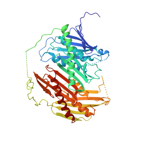
Crystal structure of Escherichia coli PNPase: central channel residues are involved in processive RNA degradation.
Shi, Z., Yang, W.Z., Lin-Chao, S., Chak, K.F., Yuan, H.S.(2008) RNA 14: 2361-2371
- PubMed: 18812438 Search on PubMedSearch on PubMed Central
- DOI: https://doi.org/10.1261/rna.1244308
- Primary Citation Related Structures:
3CDI, 3CDJ - PubMed Abstract:
Bacterial polynucleotide phosphorylase (PNPase) plays a major role in mRNA turnover by the degradation of RNA from the 3'- to 5'-ends. Here, we determined the crystal structures of the wild-type and a C-terminal KH/S1 domain-truncated mutant (DeltaKH/S1) of Escherichia coli PNPase at resolutions of 2.6 A and 2.8 A, respectively. The six RNase PH domains of the trimeric PNPase assemble into a ring-like structure containing a central channel. The truncated mutant DeltaKH/S1 bound and cleaved RNA less efficiently with an eightfold reduced binding affinity. Thermal melting and acid-induced trimer dissociation studies, analyzed by circular dichroism and dynamic light scattering, further showed that DeltaKH/S1 formed a less stable trimer than the full-length PNPase. The crystal structure of DeltaKH/S1 is more expanded, containing a slightly wider central channel than that of the wild-type PNPase, suggesting that the KH/S1 domain helps PNPase to assemble into a more compact trimer, and it regulates the channel size allosterically. Moreover, site-directed mutagenesis of several arginine residues in the channel neck regions produced defective PNPases that either bound and cleaved RNA less efficiently or generated longer cleaved oligonucleotide products, indicating that these arginines were involved in RNA binding and processive degradation. Taking these results together, we conclude that the constricted central channel and the basic-charged residues in the channel necks of PNPase play crucial roles in trapping RNA for processive exonucleolytic degradation.
- Institute of Molecular Biology, Academia Sinica, Taipei, Taiwan, Republic of China.
Organizational Affiliation: