3C5J | pdb_00003c5j

Crystal structure of HLA DR52c


Experimental Data Snapshot

  • Method: X-RAY DIFFRACTION
  • Resolution: 1.80 Å
  • R-Value Free: 
    0.227 (Depositor), 0.230 (DCC) 
  • R-Value Work: 
    0.202 (Depositor), 0.204 (DCC) 

wwPDB Validation   3D Report Full Report


Ligand Structure Quality Assessment 

Created with Raphaël 2.3.0Worse 01 BetterLigand structure goodness of fit to experimental dataBest fitted NAGClick on this verticalbar to view details

This is version 1.4 of the entry. See complete history


Literature

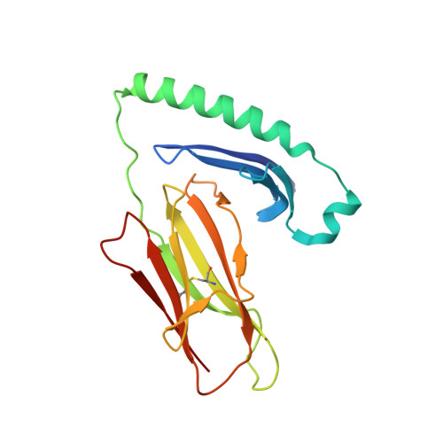
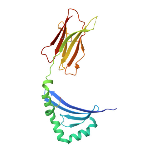
The structure of HLA-DR52c: comparison to other HLA-DRB3 alleles.

Dai, S.Crawford, F.Marrack, P.Kappler, J.W.

(2008) Proc Natl Acad Sci U S A 105: 11893-11897

  • DOI: https://doi.org/10.1073/pnas.0805810105
  • Primary Citation of Related Structures:  
    3C5J

  • PubMed Abstract: 

    Class II major histocompatibility complex (MHCII) molecules present antigens to CD4(+) T cells. In addition to the most commonly studied human MHCII isotype, HLA-DR, whose beta chain is encoded by the HLA-DRB1 locus, several other isotypes that use the same alpha chain but have beta chains encoded by other genes. These other DR molecules also are expressed in antigen-presenting cells and are known to participate in peptide presentation to T cells and to be recognized as alloantigens by other T cells. Like some of the HLA-DRB1 alleles, several of these alternate DR molecules have been associated with specific autoimmune diseases and T cell hypersensitivity. Here we present the structure of an HLA-DR molecule (DR52c) containing one of these alternate beta chains (HLA-DRB3*0301) bound to a self-peptide derived from the Tu elongation factor. The molecule shares structurally conserved elements with other MHC class II molecules but has some unique features in the peptide-binding groove. Comparison of the three major HLA-DBR3 alleles (DR52a, b, and c) suggests that they were derived from one another by recombination events that scrambled the four major peptide-binding pockets at peptide positions 1, 4, 6, and 9 but left virtually no polymorphisms elsewhere in the molecules.


  • Organizational Affiliation

    Integrated Department of Immunology, Howard Hughes Medical Institute, National Jewish Medical and Research Center, Denver, CO 80206, USA.


Macromolecules
Find similar proteins by:  (by identity cutoff)  |  3D Structure
Entity ID: 1
MoleculeChains Sequence LengthOrganismDetailsImage
HLA class II histocompatibility antigen, DR alpha chain181Homo sapiensMutation(s): 0 
Gene Names: HLA-DRAHLA-DRA1
UniProt & NIH Common Fund Data Resources
Find proteins for P01903 (Homo sapiens)
Explore P01903 
Go to UniProtKB:  P01903
PHAROS:  P01903
GTEx:  ENSG00000204287 
Entity Groups  
Sequence Clusters30% Identity50% Identity70% Identity90% Identity95% Identity100% Identity
UniProt GroupP01903
Glycosylation
Glycosylation Sites: 1Go to GlyGen: P01903-1
Sequence Annotations
Expand
  • Reference Sequence
Find similar proteins by:  (by identity cutoff)  |  3D Structure
Entity ID: 2
MoleculeChains Sequence LengthOrganismDetailsImage
MHC class II antigen190Homo sapiensMutation(s): 0 
Gene Names: HLA-DRB3
UniProt
Find proteins for B8YAC7 (Homo sapiens)
Explore B8YAC7 
Go to UniProtKB:  B8YAC7
Entity Groups  
Sequence Clusters30% Identity50% Identity70% Identity90% Identity95% Identity100% Identity
UniProt GroupB8YAC7
Glycosylation
Glycosylation Sites: 1
Sequence Annotations
Expand
  • Reference Sequence

Find similar proteins by:  Sequence   |   3D Structure  

Entity ID: 3
MoleculeChains Sequence LengthOrganismDetailsImage
Elongation factor 1-alpha 213Homo sapiensMutation(s): 0 
Gene Names: EEF1A2EEF1ALSTN
UniProt & NIH Common Fund Data Resources
Find proteins for Q05639 (Homo sapiens)
Explore Q05639 
Go to UniProtKB:  Q05639
PHAROS:  Q05639
GTEx:  ENSG00000101210 
Entity Groups  
Sequence Clusters30% Identity50% Identity70% Identity90% Identity95% Identity100% Identity
UniProt GroupQ05639
Sequence Annotations
Expand
  • Reference Sequence
Experimental Data & Validation

Experimental Data

  • Method: X-RAY DIFFRACTION
  • Resolution: 1.80 Å
  • R-Value Free:  0.227 (Depositor), 0.230 (DCC) 
  • R-Value Work:  0.202 (Depositor), 0.204 (DCC) 
Space Group: P 43 2 2
Unit Cell:
Length ( Å )Angle ( ˚ )
a = 83.38α = 90
b = 83.38β = 90
c = 132.91γ = 90
Software Package:
Software NamePurpose
DENZOdata reduction
SCALEPACKdata scaling
CNSrefinement
PDB_EXTRACTdata extraction
HKL-2000data collection
HKL-2000data reduction
HKL-2000data scaling
AMoREphasing

Structure Validation

View Full Validation Report


Ligand Structure Quality Assessment 

Created with Raphaël 2.3.0Worse 01 BetterLigand structure goodness of fit to experimental dataBest fitted NAGClick on this verticalbar to view details

Entry History 

Deposition Data

Revision History  (Full details and data files)

  • Version 1.0: 2008-08-05
    Type: Initial release
  • Version 1.1: 2011-07-13
    Changes: Non-polymer description, Version format compliance
  • Version 1.2: 2017-10-25
    Changes: Refinement description
  • Version 1.3: 2020-07-29
    Type: Remediation
    Reason: Carbohydrate remediation
    Changes: Data collection, Derived calculations, Structure summary
  • Version 1.4: 2024-10-30
    Changes: Data collection, Database references, Structure summary