3C59 | pdb_00003c59

Crystal structure of the ligand-bound glucagon-like peptide-1 receptor extracellular domain


Experimental Data Snapshot

  • Method: X-RAY DIFFRACTION
  • Resolution: 2.30 Å
  • R-Value Free: 
    0.241 (Depositor), 0.235 (DCC) 
  • R-Value Work: 
    0.196 (Depositor), 0.194 (DCC) 
  • R-Value Observed: 
    0.199 (Depositor) 

wwPDB Validation 3D Report Full Report

Validation slider image for 3C59

Ligand Structure Quality Assessment 


This is version 1.3 of the entry. See complete history

Literature

Crystal Structure of the Ligand-bound Glucagon-like Peptide-1 Receptor Extracellular Domain

Runge, S.Thogersen, H.Madsen, K.Lau, J.Rudolph, R.

(2008) J Biological Chem 283: 11340-11347

  • DOI: https://doi.org/10.1074/jbc.M708740200
  • Primary Citation Related Structures: 
    3C59, 3C5T

  • PubMed Abstract: 

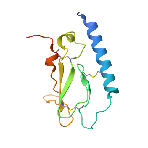
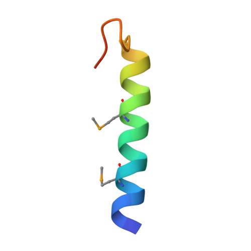
    The glucagon-like peptide-1 receptor (GLP-1R) belongs to Family B1 of the seven-transmembrane G protein-coupled receptors, and its natural agonist ligand is the peptide hormone glucagon-like peptide-1 (GLP-1). GLP-1 is involved in glucose homeostasis, and activation of GLP-1R in the plasma membrane of pancreatic beta-cells potentiates glucose-dependent insulin secretion. The N-terminal extracellular domain (nGLP-1R) is an important ligand binding domain that binds GLP-1 and the homologous peptide Exendin-4 with differential affinity. Exendin-4 has a C-terminal extension of nine amino acid residues known as the "Trp cage", which is absent in GLP-1. The Trp cage was believed to interact with nGLP-1R and thereby explain the superior affinity of Exendin-4. However, the molecular details that govern ligand binding and specificity of nGLP-1R remain undefined. Here we report the crystal structure of human nGLP-1R in complex with the antagonist Exendin-4(9-39) solved by the multiwavelength anomalous dispersion method to 2.2A resolution. The structure reveals that Exendin-4(9-39) is an amphipathic alpha-helix forming both hydrophobic and hydrophilic interactions with nGLP-1R. The Trp cage of Exendin-4 is not involved in binding to nGLP-1R. The hydrophobic binding site of nGLP-1R is defined by discontinuous segments including primarily a well defined alpha-helix in the N terminus of nGLP-1R and a loop between two antiparallel beta-strands. The structure provides for the first time detailed molecular insight into ligand binding of the human GLP-1 receptor, an established target for treatment of type 2 diabetes.


  • Organizational Affiliation
    • Department of Structure and Biophysical Chemistry, Novo Nordisk, 2760 Måløv, Denmark. sffr@novonordisk.com

Macromolecule Content 

  • Total Structure Weight: 18.31 kDa 
  • Atom Count: 1,197 
  • Modeled Residue Count: 130 
  • Deposited Residue Count: 153 
  • Unique protein chains: 2

Macromolecules

Find similar proteins by:|  3D Structure
Entity ID: 1
MoleculeChains  Sequence LengthOrganismDetailsImage
Glucagon-like peptide 1 receptor122Homo sapiensMutation(s): 0 
Gene Names: Glucagon-like peptide-1 receptor(GLP1R)
Membrane Entity: Yes 
UniProt & NIH Common Fund Data Resources
Find proteins for P43220 (Homo sapiens)
Explore P43220 
Go to UniProtKB:  P43220
PHAROS:  P43220
GTEx:  ENSG00000112164 
Entity Groups
Sequence Clusters30% Identity50% Identity70% Identity90% Identity95% Identity100% Identity
UniProt GroupP43220
Sequence Annotations
Expand
Reference Sequence
Find similar proteins by:|  3D Structure
Entity ID: 2
MoleculeChains  Sequence LengthOrganismDetailsImage
Exendin-431N/AMutation(s): 1 
Membrane Entity: Yes 
UniProt
Find proteins for P26349 (Heloderma suspectum)
Explore P26349 
Go to UniProtKB:  P26349
Entity Groups
Sequence Clusters30% Identity50% Identity70% Identity90% Identity95% Identity100% Identity
UniProt GroupP26349
Sequence Annotations
Expand
Reference Sequence

Small Molecules

Ligands 1 Unique
IDChains Name / Formula / InChI Key2D Diagram3D Interactions
10M

Query on 10M


Download:Ideal Coordinates CCD File
C [auth A]decyl 4-O-alpha-D-glucopyranosyl-1-thio-beta-D-glucopyranoside
C22 H42 O10 S
YZNNXXWNKQOETJ-HYLFJBCQSA-N
Modified Residues  1 Unique
IDChains TypeFormula2D DiagramParent
MSE
Query on MSE
B
L-PEPTIDE LINKINGC5 H11 N O2 SeMET

Experimental Data & Validation

Experimental Data

  • Method: X-RAY DIFFRACTION
  • Resolution: 2.30 Å
  • R-Value Free:  0.241 (Depositor), 0.235 (DCC) 
  • R-Value Work:  0.196 (Depositor), 0.194 (DCC) 
  • R-Value Observed: 0.199 (Depositor) 
Space Group: P 31 2 1
Unit Cell:
Length ( Å )Angle ( ˚ )
a = 75.85α = 90
b = 75.85β = 90
c = 87.55γ = 120
Software Package:
Software NamePurpose
REFMACrefinement
XDSdata scaling
XDSdata reduction
XSCALEdata scaling
autoSHARPphasing

Structure Validation

View Full Validation Report


Ligand Structure Quality Assessment 


Entry History 

Deposition Data

  • Released Date: 2008-02-19 
  • Deposition Author(s): Runge, S.

Revision History  (Full details and data files)

  • Version 1.0: 2008-02-19
    Type: Initial release
  • Version 1.1: 2011-07-13
    Changes: Advisory, Version format compliance
  • Version 1.2: 2018-05-23
    Changes: Data collection
  • Version 1.3: 2024-11-20
    Changes: Data collection, Database references, Derived calculations, Structure summary