3C2J | pdb_00003c2j

Crystal structure analysis of trioxacarcin A covalently bound to d(AACCGGTT)

  • Classification: DNA
  • Mutation(s): No 

  • Deposited: 2008-01-25 Released: 2008-06-03 
  • Deposition Author(s): Pfoh, R., Sheldrick, G.M.

Experimental Data Snapshot

  • Method: X-RAY DIFFRACTION
  • Resolution: 1.78 Å
  • R-Value Free: 
    0.265 (Depositor), 0.268 (DCC) 
  • R-Value Work: 
    0.220 (Depositor), 0.236 (DCC) 
  • R-Value Observed: 
    0.222 (Depositor) 

wwPDB Validation 3D Report Full Report

Validation slider image for 3C2J

Ligand Structure Quality Assessment 


This is version 1.2 of the entry. See complete history

Literature

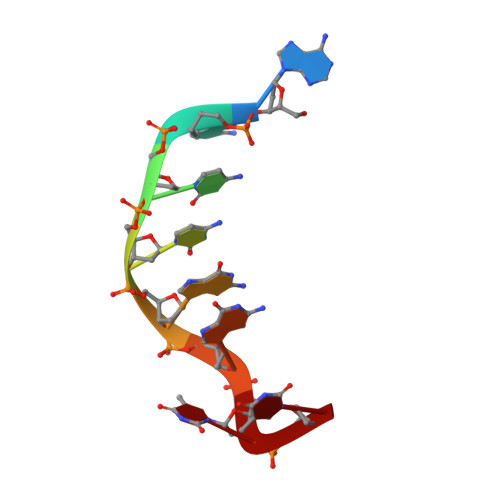
Crystal structure of trioxacarcin A covalently bound to DNA

Pfoh, R.Laatsch, H.Sheldrick, G.M.

(2008) Nucleic Acids Res 36: 3508-3514

  • DOI: https://doi.org/10.1093/nar/gkn245
  • Primary Citation Related Structures: 
    3C2J

  • PubMed Abstract: 

    We report a crystal structure that shows an antibiotic that extracts a nucleobase from a DNA molecule 'caught in the act' after forming a covalent bond but before departing with the base. The structure of trioxacarcin A covalently bound to double-stranded d(AACCGGTT) was determined to 1.78 A resolution by MAD phasing employing brominated oligonucleotides. The DNA-drug complex has a unique structure that combines alkylation (at the N7 position of a guanine), intercalation (on the 3'-side of the alkylated guanine), and base flip-out. An antibiotic-induced flipping-out of a single, nonterminal nucleobase from a DNA duplex was observed for the first time in a crystal structure.


  • Organizational Affiliation
    • Lehrstuhl für Strukturchemie, Georg-August-Universität, Tammannstr. 4, 37077 Göttingen, Germany.

Macromolecule Content 

  • Total Structure Weight: 6.61 kDa 
  • Atom Count: 467 
  • Modeled Residue Count: 16 
  • Deposited Residue Count: 16 
  • Unique nucleic acid chains: 1

Macromolecules

Find similar nucleic acids by:  Sequence
Entity ID: 1
MoleculeChains LengthOrganismImage
DNA (5'-D(*DAP*DAP*DCP*DCP*DGP*DGP*DTP*DT)-3')
A, B
8N/A
Sequence Annotations
Expand
Reference Sequence

Experimental Data & Validation

Experimental Data

  • Method: X-RAY DIFFRACTION
  • Resolution: 1.78 Å
  • R-Value Free:  0.265 (Depositor), 0.268 (DCC) 
  • R-Value Work:  0.220 (Depositor), 0.236 (DCC) 
  • R-Value Observed: 0.222 (Depositor) 
Space Group: P 41 2 2
Unit Cell:
Length ( Å )Angle ( ˚ )
a = 37.6α = 90
b = 37.6β = 90
c = 91.62γ = 90
Software Package:
Software NamePurpose
REFMACrefinement
PDB_EXTRACTdata extraction
MAR345dtbdata collection
HKL-2000data reduction
SADABSdata scaling
SHELXDEphasing

Structure Validation

View Full Validation Report


Ligand Structure Quality Assessment 


Entry History 

Deposition Data

Revision History  (Full details and data files)

  • Version 1.0: 2008-06-03
    Type: Initial release
  • Version 1.1: 2011-07-13
    Changes: Advisory, Refinement description, Version format compliance
  • Version 1.2: 2024-03-13
    Changes: Data collection, Database references, Derived calculations