Short strong hydrogen bonds in proteins: a case study of rhamnogalacturonan acetylesterase
Langkilde, A., Kristensen, S.M., Lo Leggio, L., Jensen, J.H., Houk, A.R., Navarro Poulsen, J.C., Kauppinen, S., Larsen, S.(2008) Acta Crystallogr D Biol Crystallogr 64: 851-863
- PubMed: 18645234 Search on PubMedSearch on PubMed Central
- DOI: https://doi.org/10.1107/S0907444908017083
- Primary Citation Related Structures:
3C1U - PubMed Abstract:
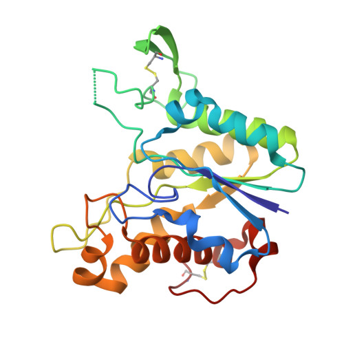
An extremely low-field signal (at approximately 18 p.p.m.) in the (1)H NMR spectrum of rhamnogalacturonan acetylesterase (RGAE) shows the presence of a short strong hydrogen bond in the structure. This signal was also present in the mutant RGAE D192N, in which Asp192, which is part of the catalytic triad, has been replaced with Asn. A careful analysis of wild-type RGAE and RGAE D192N was conducted with the purpose of identifying possible candidates for the short hydrogen bond with the 18 p.p.m. deshielded proton. Theoretical calculations of chemical shift values were used in the interpretation of the experimental (1)H NMR spectra. The crystal structure of RGAE D192N was determined to 1.33 A resolution and refined to an R value of 11.6% for all data. The structure is virtually identical to the high-resolution (1.12 A) structure of the wild-type enzyme except for the interactions involving the mutation and a disordered loop. Searches of the Cambridge Structural Database were conducted to obtain information on the donor-acceptor distances of different types of hydrogen bonds. The short hydrogen-bond interactions found in RGAE have equivalents in small-molecule structures. An examination of the short hydrogen bonds in RGAE, the calculated pK(a) values and solvent-accessibilities identified a buried carboxylic acid carboxylate hydrogen bond between Asp75 and Asp87 as the likely origin of the 18 p.p.m. signal. Similar hydrogen-bond interactions between two Asp or Glu carboxy groups were found in 16% of a homology-reduced set of high-quality structures extracted from the PDB. The shortest hydrogen bonds in RGAE are all located close to the active site and short interactions between Ser and Thr side-chain OH groups and backbone carbonyl O atoms seem to play an important role in the stability of the protein structure. These results illustrate the significance of short strong hydrogen bonds in proteins.
- Department of Chemistry, University of Copenhagen, Universitetsparken 5, DK-2100 Copenhagen, Denmark.
Organizational Affiliation: