3C0J | pdb_00003c0j

Structure of E. coli dihydrodipicolinate synthase complexed with hydroxypyruvate


Experimental Data Snapshot

  • Method: X-RAY DIFFRACTION
  • Resolution: 2.40 Å
  • R-Value Free: 
    0.237 (Depositor), 0.243 (DCC) 
  • R-Value Work: 
    0.170 (Depositor), 0.178 (DCC) 
  • R-Value Observed: 
    0.173 (Depositor) 

Starting Model: experimental
View more details

wwPDB Validation 3D Report Full Report

Validation slider image for 3C0J

This is version 1.5 of the entry. See complete history

Literature

Conserved main-chain peptide distortions: A proposed role for Ile203 in catalysis by dihydrodipicolinate synthase

Dobson, R.C.J.Griffin, M.D.W.Devenish, S.R.A.Pearce, F.G.Hutton, C.A.Gerrard, J.A.Jameson, G.B.Perugini, M.A.

(2008) Protein Sci 17: 2080-2090

  • DOI: https://doi.org/10.1110/ps.037440.108
  • Primary Citation Related Structures: 
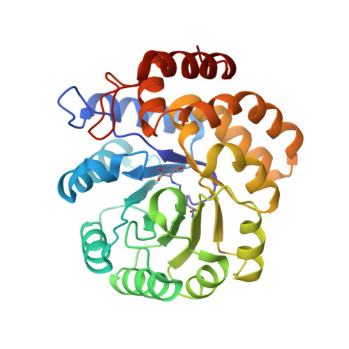
    3C0J

  • PubMed Abstract: 

    In recent years, dihydrodipicolinate synthase (DHDPS, E.C. 4.2.1.52) has received considerable attention from a mechanistic and structural viewpoint. DHDPS catalyzes the reaction of (S)-aspartate-beta-semialdehyde with pyruvate, which is bound via a Schiff base to a conserved active-site lysine (Lys161 in the enzyme from Escherichia coli). To probe the mechanism of DHDPS, we have studied the inhibition of E. coli DHDPS by the substrate analog, beta-hydroxypyruvate. The K (i) was determined to be 0.21 (+/-0.02) mM, similar to that of the allosteric inhibitor, (S)-lysine, and beta-hydroxypyruvate was observed to cause time-dependent inhibition. The inhibitory reaction with beta-hydroxypyruvate could be qualitatively followed by mass spectrometry, which showed initial noncovalent adduct formation, followed by the slow formation of the covalent adduct. It is unclear whether beta-hydroxypyruvate plays a role in regulating the biosynthesis of meso-diaminopimelate and (S)-lysine in E. coli, although we note that it is present in vivo. The crystal structure of DHDPS complexed with beta-hydroxypyruvate was solved. The active site clearly showed the presence of the inhibitor covalently bound to the Lys161. Interestingly, the hydroxyl group of beta-hydroxypyruvate was hydrogen-bonded to the main-chain carbonyl of Ile203. This provides insight into the possible catalytic role played by this peptide unit, which has a highly strained torsion angle (omega approximately 201 degrees ). A survey of the known DHDPS structures from other organisms shows this distortion to be a highly conserved feature of the DHDPS active site, and we propose that this peptide unit plays a critical role in catalysis.


  • Organizational Affiliation
    • Department of Biochemistry and Molecular Biology, University of Melbourne, Parkville, Victoria 3010, Australia. rdobson@unimelb.edu.au

Macromolecule Content 

  • Total Structure Weight: 63.32 kDa 
  • Atom Count: 4,897 
  • Modeled Residue Count: 584 
  • Deposited Residue Count: 584 
  • Unique protein chains: 1

Macromolecules

Find similar proteins by:|  3D Structure
Entity ID: 1
MoleculeChains  Sequence LengthOrganismDetailsImage
Dihydrodipicolinate synthase
A, B
292Escherichia coli K-12Mutation(s): 0 
Gene Names: dapA
EC: 4.2.1.52 (PDB Primary Data), 4.3.3.7 (UniProt)
UniProt
Find proteins for P0A6L2 (Escherichia coli (strain K12))
Explore P0A6L2 
Go to UniProtKB:  P0A6L2
Entity Groups
Sequence Clusters30% Identity50% Identity70% Identity90% Identity95% Identity100% Identity
UniProt GroupP0A6L2
Sequence Annotations
Expand
Reference Sequence

Small Molecules

Ligands 3 Unique
IDChains Name / Formula / InChI Key2D Diagram3D Interactions
PO4

Query on PO4


Download:Ideal Coordinates CCD File
H [auth B],
I [auth B]
PHOSPHATE ION
O4 P
NBIIXXVUZAFLBC-UHFFFAOYSA-K
GOL

Query on GOL


Download:Ideal Coordinates CCD File
D [auth A],
E [auth A],
F [auth B]
GLYCEROL
C3 H8 O3
PEDCQBHIVMGVHV-UHFFFAOYSA-N
K

Query on K


Download:Ideal Coordinates CCD File
C [auth A],
G [auth B]
POTASSIUM ION
K
NPYPAHLBTDXSSS-UHFFFAOYSA-N
Modified Residues  1 Unique
IDChains TypeFormula2D DiagramParent
KYQ
Query on KYQ
A, B
L-PEPTIDE LINKINGC9 H16 N2 O5LYS

Experimental Data & Validation

Experimental Data

  • Method: X-RAY DIFFRACTION
  • Resolution: 2.40 Å
  • R-Value Free:  0.237 (Depositor), 0.243 (DCC) 
  • R-Value Work:  0.170 (Depositor), 0.178 (DCC) 
  • R-Value Observed: 0.173 (Depositor) 
Space Group: P 31 2 1
Unit Cell:
Length ( Å )Angle ( ˚ )
a = 120.967α = 90
b = 120.967β = 90
c = 110.639γ = 120
Software Package:
Software NamePurpose
REFMACrefinement
CrystalCleardata collection
d*TREKdata reduction
d*TREKdata scaling
AMoREphasing

Structure Validation

View Full Validation Report


Entry History 

Deposition Data

Revision History  (Full details and data files)

  • Version 1.0: 2008-09-23
    Type: Initial release
  • Version 1.1: 2011-07-13
    Changes: Non-polymer description, Version format compliance
  • Version 1.2: 2018-01-31
    Changes: Experimental preparation
  • Version 1.3: 2023-11-01
    Changes: Data collection, Database references, Derived calculations, Refinement description
  • Version 1.4: 2023-11-15
    Changes: Data collection
  • Version 1.5: 2024-10-30
    Changes: Structure summary