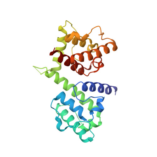
High-resolution cryo-EM structure of the F-actin-fimbrin/plastin ABD2 complex.
Galkin, V.E., Orlova, A., Cherepanova, O., Lebart, M.C., Egelman, E.H.(2008) Proc Natl Acad Sci U S A 105: 1494-1498
- PubMed: 18234857
- DOI: https://doi.org/10.1073/pnas.0708667105
- Primary Citation of Related Structures:
3BYH - PubMed Abstract:
Many actin binding proteins have a modular architecture, and calponin-homology (CH) domains are one such structurally conserved module found in numerous proteins that interact with F-actin. The manner in which CH-domains bind F-actin has been controversial. Using cryo-EM and a single-particle approach to helical reconstruction, we have generated 12-A-resolution maps of F-actin alone and F-actin decorated with a fragment of human fimbrin (L-plastin) containing tandem CH-domains. The high resolution allows an unambiguous fit of the crystal structure of fimbrin into the map. The interaction between fimbrin ABD2 (actin binding domain 2) and F-actin is different from any interaction previously observed or proposed for tandem CH-domain proteins, showing that the structural conservation of the CH-domains does not lead to a conserved mode of interaction with F-actin. Both the stapling of adjacent actin protomers and the additional closure of the nucleotide binding cleft in F-actin when the fimbrin fragment binds may explain how fimbrin can stabilize actin filaments. A mechanism is proposed where ABD1 of fimbrin becomes activated for binding a second actin filament after ABD2 is bound to a first filament, and this can explain how mutations of residues buried in the interface between ABD2 and ABD1 can rescue temperature-sensitive defects in actin.
- Department of Biochemistry and Molecular Genetics, University of Virginia, Charlottesville, VA 22908-0733, USA.
Organizational Affiliation:


