3BSD | pdb_00003bsd

Light harvesting protein from RC of Chlorobium tepidum


Experimental Data Snapshot

  • Method: X-RAY DIFFRACTION
  • Resolution: 2.30 Å
  • R-Value Free: 
    0.273 (Depositor) 
  • R-Value Work: 
    0.174 (Depositor), 0.181 (DCC) 
  • R-Value Observed: 
    0.178 (Depositor) 

Starting Model: experimental
View more details

wwPDB Validation 3D Report Full Report

Validation slider image for 3BSD

Ligand Structure Quality Assessment 


This is version 1.3 of the entry. See complete history

Literature

Evolution of photosystem I - from symmetry through pseudo-symmetry to asymmetry.

Ben-Shem, A.Frolow, F.Nelson, N.

(2004) FEBS Lett 564: 274-280

  • DOI: https://doi.org/10.1016/S0014-5793(04)00360-6
  • Primary Citation Related Structures: 
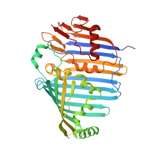
    3BSD

  • PubMed Abstract: 

    The evolution of photosystem (PS) I was probably initiated by the formation of a homodimeric reaction center similar to the one currently present in green bacteria. Gene duplication has generated a heterodimeric reaction center that subsequently evolved to the PSI present in cyanobacteria, algae and plant chloroplasts. During the evolution of PSI several attempts to maximize the efficiency of light harvesting took place in the various organisms. In the Chlorobiaceae, chlorosomes and FMO were added to the homodimeric reaction center. In cyanobacteria phycobilisomes and CP43' evolved to cope with the light limitations and stress conditions. The plant PSI utilizes a modular arrangement of membrane light-harvesting proteins (LHCI). We obtained structural information from the two ends of the evolutionary spectrum. Novel features in the structure of Chlorobium tepidum FMO are reported in this communication. Our structure of plant PSI reveals that the addition of subunit G provided the template for LHCI binding, and the addition of subunit H prevented the possibility of trimer formation and provided a binding site for LHCII and the onset of energy spillover from PSII to PSI.


  • Organizational Affiliation
    • Department of Biochemistry, The George S. Wise Faculty of Life Sciences, Tel Aviv University, Tel Aviv 69978, Israel.

Macromolecule Content 

  • Total Structure Weight: 47.66 kDa 
  • Atom Count: 3,481 
  • Modeled Residue Count: 359 
  • Deposited Residue Count: 366 
  • Unique protein chains: 1

Macromolecules

Find similar proteins by:|  3D Structure
Entity ID: 1
MoleculeChains  Sequence LengthOrganismDetailsImage
Bacteriochlorophyll a protein366Chlorobaculum tepidumMutation(s): 0 
UniProt
Find proteins for Q46393 (Chlorobaculum tepidum (strain ATCC 49652 / DSM 12025 / NBRC 103806 / TLS))
Explore Q46393 
Go to UniProtKB:  Q46393
Entity Groups
Sequence Clusters30% Identity50% Identity70% Identity90% Identity95% Identity100% Identity
UniProt GroupQ46393
Sequence Annotations
Expand
Reference Sequence

Small Molecules

Ligands 2 Unique
IDChains Name / Formula / InChI Key2D Diagram3D Interactions
BCL

Query on BCL


Download:Ideal Coordinates CCD File
C [auth A]
D [auth A]
E [auth A]
F [auth A]
G [auth A]
C [auth A],
D [auth A],
E [auth A],
F [auth A],
G [auth A],
H [auth A],
I [auth A],
J [auth A]
BACTERIOCHLOROPHYLL A
C55 H74 Mg N4 O6
DSJXIQQMORJERS-AGGZHOMASA-M
MG

Query on MG


Download:Ideal Coordinates CCD File
B [auth A]MAGNESIUM ION
Mg
JLVVSXFLKOJNIY-UHFFFAOYSA-N

Experimental Data & Validation

Experimental Data

  • Method: X-RAY DIFFRACTION
  • Resolution: 2.30 Å
  • R-Value Free:  0.273 (Depositor) 
  • R-Value Work:  0.174 (Depositor), 0.181 (DCC) 
  • R-Value Observed: 0.178 (Depositor) 
Space Group: I 41 3 2
Unit Cell:
Length ( Å )Angle ( ˚ )
a = 167.695α = 90
b = 167.695β = 90
c = 167.695γ = 90
Software Package:
Software NamePurpose
REFMACrefinement
DENZOdata reduction
SCALEPACKdata scaling
AMoREphasing

Structure Validation

View Full Validation Report


Ligand Structure Quality Assessment 


Entry History 

Deposition Data

Revision History  (Full details and data files)

  • Version 1.0: 2008-01-22
    Type: Initial release
  • Version 1.1: 2011-07-13
    Changes: Advisory, Version format compliance
  • Version 1.2: 2017-10-25
    Changes: Refinement description
  • Version 1.3: 2023-11-01
    Changes: Data collection, Database references, Derived calculations, Refinement description