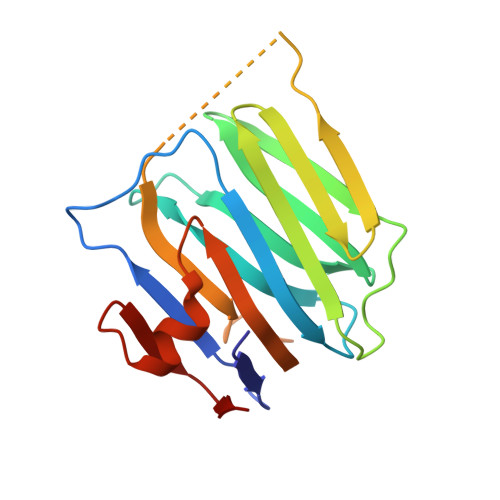
3BOP | pdb_00003bop

Structure of mouse beta-neurexin 2D4


Experimental Data Snapshot

  • Method: X-RAY DIFFRACTION
  • Resolution: 3.00 Å
  • R-Value Free: 
    0.289 (Depositor), 0.224 (DCC) 
  • R-Value Work: 
    0.211 (Depositor), 0.218 (DCC) 
  • R-Value Observed: 
    0.215 (Depositor) 

wwPDB Validation 3D Report Full Report

Validation slider image for 3BOP

This is version 1.2 of the entry. See complete history. 

Literature

Crystal Structures of beta-Neurexin 1 and beta-Neurexin 2 Ectodomains and Dynamics of Splice Insertion Sequence 4.

Koehnke, J., Jin, X., Trbovic, N., Katsamba, P.S., Brasch, J., Ahlsen, G., Scheiffele, P., Honig, B., Palmer, A.G., Shapiro, L.

(2008) Structure 16: 410-421

  • DOI: https://doi.org/10.1016/j.str.2007.12.024
  • Primary Citation Related Structures: 
    3BOD, 3BOP

  • PubMed Abstract: 

    Presynaptic neurexins (NRXs) bind to postsynaptic neuroligins (NLs) to form Ca(2+)-dependent complexes that bridge neural synapses. beta-NRXs bind NLs through their LNS domains, which contain a single site of alternative splicing (splice site 4) giving rise to two isoforms: +4 and Delta. We present crystal structures of the Delta isoforms of the LNS domains from beta-NRX1 and beta-NRX2, crystallized in the presence of Ca(2+) ions. The Ca(2+)-binding site is disordered in the beta-NRX2 structure, but the 1.7 A beta-NRX1 structure reveals a single Ca(2+) ion, approximately 12 A from the splice insertion site, with one coordinating ligand donated by a glutamic acid from an adjacent beta-NRX1 molecule. NMR studies of beta-NRX1+4 show that the insertion sequence is unstructured, and remains at least partially disordered in complex with NL. These results raise the possibility that beta-NRX insertion sequence 4 may function in roles independent of neuroligin binding.


  • Organizational Affiliation: 
    • Department of Biochemistry and Molecular Biophysics, Columbia University, 630 West 168th Street, New York, NY 10032, USA.

Macromolecule Content 

  • Total Structure Weight: 57.12 kDa 
  • Atom Count: 4,009 
  • Modeled Residue Count: 505 
  • Deposited Residue Count: 525 
  • Unique protein chains: 1

Macromolecules

Find similar proteins by:|  3D Structure
Entity ID: 1
MoleculeChains  Sequence LengthOrganismDetailsImage
beta-Neurexin 2D4
A, B, C
175Mus musculusMutation(s): 0 
Gene Names: Nrxn2
UniProt
Find proteins for Q3TQ54 (Mus musculus)
Explore Q3TQ54 
Go to UniProtKB:  Q3TQ54
Entity Groups
Sequence Clusters30% Identity50% Identity70% Identity90% Identity95% Identity100% Identity
UniProt GroupQ3TQ54
Sequence Annotations
Expand
Reference Sequence

Experimental Data & Validation

Experimental Data

  • Method: X-RAY DIFFRACTION
  • Resolution: 3.00 Å
  • R-Value Free:  0.289 (Depositor), 0.224 (DCC) 
  • R-Value Work:  0.211 (Depositor), 0.218 (DCC) 
  • R-Value Observed: 0.215 (Depositor) 
Space Group: C 1 2 1
Unit Cell:
Length ( Å )Angle ( ˚ )
a = 95.485α = 90
b = 96.983β = 105.98
c = 62.584γ = 90
Software Package:
Software NamePurpose
REFMACrefinement
PDB_EXTRACTdata extraction
ADSCdata collection
DENZOdata reduction
SCALEPACKdata scaling
PHASERphasing

Structure Validation

View Full Validation Report


Entry History 

Deposition Data

Revision History  (Full details and data files)

  • Version 1.0: 2008-03-25
    Type: Initial release
  • Version 1.1: 2011-07-13
    Changes: Advisory, Version format compliance
  • Version 1.2: 2024-02-21
    Changes: Data collection, Database references