3BOA | pdb_00003boa

Crystal structure of yeast protein disulfide isomerase.


Experimental Data Snapshot

  • Method: X-RAY DIFFRACTION
  • Resolution: 3.70 Å
  • R-Value Free: 
    0.340 (Depositor), 0.239 (DCC) 
  • R-Value Work: 
    0.239 (Depositor), 0.246 (DCC) 
  • R-Value Observed: 
    0.244 (Depositor) 

Starting Model: experimental
View more details

wwPDB Validation 3D Report Full Report

Validation slider image for 3BOA

This is version 1.3 of the entry. See complete history

Literature

The Catalytic Activity of Protein-disulfide Isomerase Requires a Conformationally Flexible Molecule.

Tian, G.Kober, F.X.Lewandrowski, U.Sickmann, A.Lennarz, W.J.Schindelin, H.

(2008) J Biological Chem 283: 33630-33640

  • DOI: https://doi.org/10.1074/jbc.M806026200
  • Primary Citation Related Structures: 
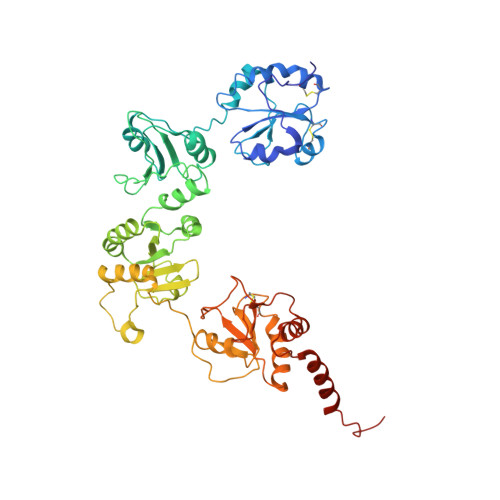
    3BOA

  • PubMed Abstract: 

    Protein-disulfide isomerase (PDI) catalyzes the formation of the correct pattern of disulfide bonds in secretory proteins. A low resolution crystal structure of yeast PDI described here reveals large scale conformational changes compared with the initially reported structure, indicating that PDI is a highly flexible molecule with its catalytic domains, a and a', representing two mobile arms connected to a more rigid core composed of the b and b' domains. Limited proteolysis revealed that the linker between the a domain and the core is more susceptible to degradation than that connecting the a' domain to the core. By restricting the two arms with inter-domain disulfide bonds, the molecular flexibility of PDI, especially that of its a domain, was demonstrated to be essential for the enzymatic activity in vitro and in vivo. The crystal structure also featured a PDI dimer, and a propensity to dimerize in solution and in the ER was confirmed by cross-linking experiments and the split green fluorescent protein system. Although sedimentation studies suggested that the self-association of PDI is weak, we hypothesize that PDI exists as an interconvertible mixture of monomers and dimers in the endoplasmic reticulum due to its high abundance in this compartment.


  • Organizational Affiliation
    • Department of Biochemistry and Cell Biology, Stony Brook University, Stony Brook, New York 11794-5215, USA.

Macromolecule Content 

  • Total Structure Weight: 56.42 kDa 
  • Atom Count: 3,864 
  • Modeled Residue Count: 490 
  • Deposited Residue Count: 504 
  • Unique protein chains: 1

Macromolecules

Find similar proteins by:|  3D Structure
Entity ID: 1
MoleculeChains  Sequence LengthOrganismDetailsImage
Protein disulfide-isomerase504Saccharomyces cerevisiaeMutation(s): 0 
Gene Names: PDI1MFP1TRG1
EC: 5.3.4.1
UniProt
Find proteins for P17967 (Saccharomyces cerevisiae (strain ATCC 204508 / S288c))
Explore P17967 
Go to UniProtKB:  P17967
Entity Groups
Sequence Clusters30% Identity50% Identity70% Identity90% Identity95% Identity100% Identity
UniProt GroupP17967
Sequence Annotations
Expand
Reference Sequence

Experimental Data & Validation

Experimental Data

  • Method: X-RAY DIFFRACTION
  • Resolution: 3.70 Å
  • R-Value Free:  0.340 (Depositor), 0.239 (DCC) 
  • R-Value Work:  0.239 (Depositor), 0.246 (DCC) 
  • R-Value Observed: 0.244 (Depositor) 
Space Group: C 2 2 21
Unit Cell:
Length ( Å )Angle ( ˚ )
a = 116.921α = 90
b = 123.154β = 90
c = 75.721γ = 90
Software Package:
Software NamePurpose
REFMACrefinement
CBASSdata collection
HKL-2000data reduction
HKL-2000data scaling
PHASERphasing

Structure Validation

View Full Validation Report


Entry History 

Deposition Data

Revision History  (Full details and data files)

  • Version 1.0: 2008-09-23
    Type: Initial release
  • Version 1.1: 2011-07-13
    Changes: Advisory, Version format compliance
  • Version 1.2: 2023-08-30
    Changes: Data collection, Database references, Refinement description
  • Version 1.3: 2024-10-30
    Changes: Structure summary