3BMZ | pdb_00003bmz

Violacein biosynthetic enzyme VioE


Experimental Data Snapshot

  • Method: X-RAY DIFFRACTION
  • Resolution: 1.21 Å
  • R-Value Free: 
    0.195 (Depositor), 0.200 (DCC) 
  • R-Value Work: 
    0.168 (Depositor), 0.173 (DCC) 
  • R-Value Observed: 
    0.169 (Depositor) 

wwPDB Validation 3D Report Full Report

Validation slider image for 3BMZ

This is version 1.2 of the entry. See complete history

Literature

The Violacein Biosynthetic Enzyme VioE Shares a Fold with Lipoprotein Transporter Proteins

Ryan, K.S.Balibar, C.J.Turo, K.E.Walsh, C.T.Drennan, C.L.

(2008) J Biological Chem 283: 6467-6475

  • DOI: https://doi.org/10.1074/jbc.M708573200
  • Primary Citation Related Structures: 
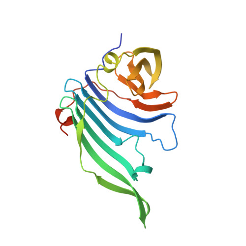
    3BMZ

  • PubMed Abstract: 

    VioE, an unusual enzyme with no characterized homologues, plays a key role in the biosynthesis of violacein, a purple pigment with antibacterial and cytotoxic properties. Without bound cofactors or metals, VioE, from the bacterium Chromobacterium violaceum, mediates a 1,2 shift of an indole ring and oxidative chemistry to generate prodeoxyviolacein, a precursor to violacein. Our 1.21 A resolution structure of VioE shows that the enzyme shares a core fold previously described for lipoprotein transporter proteins LolA and LolB. For both LolB and VioE, a bound polyethylene glycol molecule suggests the location of the binding and/or active site of the protein. Mutations of residues near the bound polyethylene glycol molecule in VioE have identified the active site and five residues important for binding or catalysis. This structural and mutagenesis study suggests that VioE acts as a catalytic chaperone, using a fold previously associated with lipoprotein transporters to catalyze the production of its prodeoxyviolacein product.


  • Organizational Affiliation
    • Department of Biology and Department of Chemistry, Massachusetts Institute of Technology, Cambridge, MA 02139, USA.

Macromolecule Content 

  • Total Structure Weight: 46.1 kDa 
  • Atom Count: 3,886 
  • Modeled Residue Count: 375 
  • Deposited Residue Count: 398 
  • Unique protein chains: 1

Macromolecules

Find similar proteins by:|  3D Structure
Entity ID: 1
MoleculeChains  Sequence LengthOrganismDetailsImage
Putative uncharacterized protein
A, B
199Chromobacterium violaceum ATCC 12472Mutation(s): 0 
Gene Names: VioE
UniProt
Find proteins for Q7NSZ5 (Chromobacterium violaceum (strain ATCC 12472 / DSM 30191 / JCM 1249 / CCUG 213 / NBRC 12614 / NCIMB 9131 / NCTC 9757 / MK))
Explore Q7NSZ5 
Go to UniProtKB:  Q7NSZ5
Entity Groups
Sequence Clusters30% Identity50% Identity70% Identity90% Identity95% Identity100% Identity
UniProt GroupQ7NSZ5
Sequence Annotations
Expand
Reference Sequence

Small Molecules

Experimental Data & Validation

Experimental Data

  • Method: X-RAY DIFFRACTION
  • Resolution: 1.21 Å
  • R-Value Free:  0.195 (Depositor), 0.200 (DCC) 
  • R-Value Work:  0.168 (Depositor), 0.173 (DCC) 
  • R-Value Observed: 0.169 (Depositor) 
Space Group: P 21 21 21
Unit Cell:
Length ( Å )Angle ( ˚ )
a = 53.527α = 90
b = 82.477β = 90
c = 90.633γ = 90
Software Package:
Software NamePurpose
REFMACrefinement
ADSCdata collection
DENZOdata reduction
SCALEPACKdata scaling
SHARPphasing

Structure Validation

View Full Validation Report


Entry History 

Deposition Data

Revision History  (Full details and data files)

  • Version 1.0: 2008-01-01
    Type: Initial release
  • Version 1.1: 2011-07-13
    Changes: Source and taxonomy, Version format compliance
  • Version 1.2: 2024-02-21
    Changes: Data collection, Database references, Derived calculations