6-Azido-7-nitro-1,4-dihydroquinoxaline-2,3-dione (ANQX) forms an irreversible bond to the active site of the GluR2 AMPA receptor.
Cruz, L.A., Estebanez-Perpina, E., Pfaff, S., Borngraeber, S., Bao, N., Blethrow, J., Fletterick, R.J., England, P.M.(2008) J Med Chem 51: 5856-5860
- PubMed: 18754610 Search on PubMedSearch on PubMed Central
- DOI: https://doi.org/10.1021/jm701517b
- Primary Citation Related Structures:
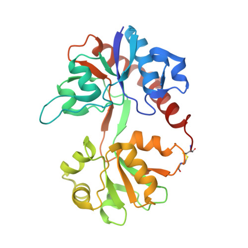
3BKI - PubMed Abstract:
AMPA receptors mediate fast excitatory synaptic transmission and are essential for synaptic plasticity. ANQX, a photoreactive AMPA receptor antagonist, is an important biological probe used to irreversibly inactivate AMPA receptors. Here, using X-ray crystallography and mass spectroscopy, we report that ANQX forms two major products in the presence of the GluR2 AMPAR ligand-binding core (S1S2J). Upon photostimulation, ANQX reacts intramolecularly to form FQX or intermolecularly to form a covalent adduct with Glu705.
- Graduate Program in Chemistry and Chemical Biology, University of California San Francisco, San Francisco, California 94158-2517, USA.
Organizational Affiliation: