3BHF | pdb_00003bhf

Crystal structure of R49K mutant of Monomeric Sarcosine Oxidase crystallized in PEG as precipitant


Experimental Data Snapshot

  • Method: X-RAY DIFFRACTION
  • Resolution: 2.10 Å
  • R-Value Free: 
    0.251 (Depositor), 0.254 (DCC) 
  • R-Value Work: 
    0.202 (Depositor), 0.203 (DCC) 
  • R-Value Observed: 
    0.202 (Depositor) 

Starting Model: experimental
View more details

wwPDB Validation 3D Report Full Report

Validation slider image for 3BHF

Ligand Structure Quality Assessment 


This is version 1.4 of the entry. See complete history

Literature

Arginine 49 is a bifunctional residue important in catalysis and biosynthesis of monomeric sarcosine oxidase: a context-sensitive model for the electrostatic impact of arginine to lysine mutations.

Hassan-Abdallah, A.Zhao, G.Chen, Z.W.Mathews, F.S.Schuman Jorns, M.

(2008) Biochemistry 47: 2913-2922

  • DOI: https://doi.org/10.1021/bi702351v
  • Primary Citation Related Structures: 
    3BHF, 3BHK

  • PubMed Abstract: 

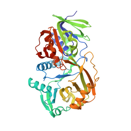
    Monomeric sarcosine oxidase (MSOX) contains covalently bound FAD and catalyzes the oxidative demethylation of sarcosine ( N-methylglycine). The side chain of Arg49 is in van der Waals contact with the si face of the flavin ring; sarcosine binds just above the re face. Covalent flavin attachment requires a basic residue (Arg or Lys) at position 49. Although flavinylation is scarcely affected, mutation of Arg49 to Lys causes a 40-fold decrease in k cat and a 150-fold decrease in k cat/ K m sarcosine. The overall structure of the Arg49Lys mutant is very similar to wild-type MSOX; the side chain of Lys49 in the mutant is nearly congruent to that of Arg49 in the wild-type enzyme. The Arg49Lys mutant exhibits several features consistent with a less electropositive active site: (1) Charge transfer bands observed for mutant enzyme complexes with competitive inhibitors absorb at higher energy than the corresponding wild-type complexes. (2) The p K a for ionization at N(3)H of FAD is more than two pH units higher in the mutant than in wild-type MSOX. (3) The reduction potential of the oxidized/radical couple in the mutant is 100 mV lower than in the wild-type enzyme. The lower reduction potential is likely to be a major cause of the reduced catalytic activity of the mutant. Electrostatic interactions with Arg49 play an important role in catalysis and covalent flavinylation. A context-sensitive model for the electrostatic impact of an arginine to lysine mutation can account for the dramatically different consequences of the Arg49Lys mutation on MSOX catalysis and holoenzyme biosysnthesis.


  • Organizational Affiliation
    • Department of Biochemistry and Molecular Biology, Drexel University College of Medicine, Philadelphia, Pennsylvania 19102, USA.

Macromolecule Content 

  • Total Structure Weight: 88.05 kDa 
  • Atom Count: 6,434 
  • Modeled Residue Count: 762 
  • Deposited Residue Count: 780 
  • Unique protein chains: 1

Macromolecules

Find similar proteins by:|  3D Structure
Entity ID: 1
MoleculeChains  Sequence LengthOrganismDetailsImage
Monomeric sarcosine oxidase
A, B
390Bacillus sp. (in: firmicutes)Mutation(s): 1 
Gene Names: soxAsox
EC: 1.5.3.1
UniProt
Find proteins for P40859 (Bacillus sp. (strain B-0618))
Explore P40859 
Go to UniProtKB:  P40859
Entity Groups
Sequence Clusters30% Identity50% Identity70% Identity90% Identity95% Identity100% Identity
UniProt GroupP40859
Sequence Annotations
Expand
Reference Sequence

Experimental Data & Validation

Experimental Data

  • Method: X-RAY DIFFRACTION
  • Resolution: 2.10 Å
  • R-Value Free:  0.251 (Depositor), 0.254 (DCC) 
  • R-Value Work:  0.202 (Depositor), 0.203 (DCC) 
  • R-Value Observed: 0.202 (Depositor) 
Space Group: P 1 21 1
Unit Cell:
Length ( Å )Angle ( ˚ )
a = 72.348α = 90
b = 70.456β = 96.77
c = 79.595γ = 90
Software Package:
Software NamePurpose
CNSrefinement
HKL-2000data collection
HKL-2000data reduction
SCALEPACKdata scaling
MOLREPphasing

Structure Validation

View Full Validation Report


Ligand Structure Quality Assessment 


Entry History 

Deposition Data

Revision History  (Full details and data files)

  • Version 1.0: 2008-02-26
    Type: Initial release
  • Version 1.1: 2011-07-13
    Changes: Version format compliance
  • Version 1.2: 2021-10-20
    Changes: Database references, Derived calculations
  • Version 1.3: 2023-08-30
    Changes: Data collection, Refinement description
  • Version 1.4: 2024-11-06
    Changes: Structure summary