


Crystal Structure for shikimate kinase from Mycobacterium tuberculosis in complex with AMP-PNP
Faim, L.M., Dias, M.V.B., Vasconcelos, I.G., Basso, L.A., Santos, D.S., Azevedo, W.F., Ruggiero, N.J.To be published.
Experimental Data Snapshot
Starting Model: experimental
View more details
Entity ID: 1 | |||||
|---|---|---|---|---|---|
| Molecule | Chains | Sequence Length | Organism | Details | Image |
| Shikimate kinase | 176 | Mycobacterium tuberculosis | Mutation(s): 0 Gene Names: aroK EC: 2.7.1.71 | 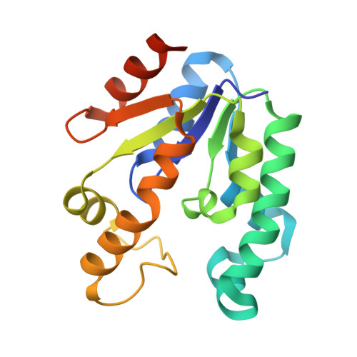 | |
UniProt | |||||
Entity Groups | |||||
| Sequence Clusters | 30% Identity50% Identity70% Identity90% Identity95% Identity100% Identity | ||||
| UniProt Group | P9WPY3 | ||||
Sequence AnnotationsExpand | |||||
Reference Sequence | |||||
| Ligands 2 Unique | |||||
|---|---|---|---|---|---|
| ID | Chains | Name / Formula / InChI Key | 2D Diagram | 3D Interactions | |
| ANP Download:Ideal Coordinates CCD File | B [auth A] | PHOSPHOAMINOPHOSPHONIC ACID-ADENYLATE ESTER C10 H17 N6 O12 P3 PVKSNHVPLWYQGJ-KQYNXXCUSA-N |  | ||
| SKM Download:Ideal Coordinates CCD File | C [auth A] | (3R,4S,5R)-3,4,5-TRIHYDROXYCYCLOHEX-1-ENE-1-CARBOXYLIC ACID C7 H10 O5 JXOHGGNKMLTUBP-HSUXUTPPSA-N |  | ||
| Length ( Å ) | Angle ( ˚ ) |
|---|---|
| a = 67.297 | α = 90 |
| b = 67.297 | β = 90 |
| c = 97.692 | γ = 120 |
| Software Name | Purpose |
|---|---|
| REFMAC | refinement |
| MAR345dtb | data collection |
| MOSFLM | data reduction |
| SCALA | data scaling |
| AMoRE | phasing |


