3B9G | pdb_00003b9g

Crystal structure of loop deletion mutant of Trypanosoma vivax nucleoside hydrolase (3GTvNH) in complex with ImmH


Experimental Data Snapshot

  • Method: X-RAY DIFFRACTION
  • Resolution: 1.40 Å
  • R-Value Free: 
    0.204 (Depositor) 
  • R-Value Work: 
    0.177 (Depositor), 0.178 (DCC) 
  • R-Value Observed: 
    0.177 (Depositor) 

Starting Model: experimental
View more details

wwPDB Validation 3D Report Full Report

Validation slider image for 3B9G

Ligand Structure Quality Assessment 


This is version 1.4 of the entry. See complete history

Literature

A Flexible Loop as a Functional Element in the Catalytic Mechanism of Nucleoside Hydrolase from Trypanosoma vivax.

Vandemeulebroucke, A.De Vos, S.Van Holsbeke, E.Steyaert, J.Versees, W.

(2008) J Biological Chem 283: 22272-22282

  • DOI: https://doi.org/10.1074/jbc.M803705200
  • Primary Citation Related Structures: 
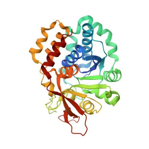
    3B9G

  • PubMed Abstract: 

    The nucleoside hydrolase of Trypanosoma vivax hydrolyzes the N-glycosidic bond of purine nucleosides. Structural and kinetic studies on this enzyme have suggested a catalytic role for a flexible loop in the vicinity of the active sites. Here we present the analysis of the role of this flexible loop via the combination of a proline scan of the loop, loop deletion mutagenesis, steady state and pre-steady state analysis, and x-ray crystallography. Our analysis reveals that this loop has an important role in leaving group activation and product release. The catalytic role involves the entire loop and could only be perturbed by deletion of the entire loop and not by single site mutagenesis. We present evidence that the loop closes over the active site during catalysis, thereby ordering a water channel that is involved in leaving group activation. Once chemistry has taken place, the loop dynamics determine the rate of product release.


  • Organizational Affiliation
    • Department of Ultrastructure, Vrije Universiteit Brussel (VIB), Pleinlaan 2, Brussels 1050, Belgium.

Macromolecule Content 

  • Total Structure Weight: 73.29 kDa 
  • Atom Count: 5,668 
  • Modeled Residue Count: 633 
  • Deposited Residue Count: 656 
  • Unique protein chains: 1

Macromolecules

Find similar proteins by:|  3D Structure
Entity ID: 1
MoleculeChains  Sequence LengthOrganismDetailsImage
IAG-nucleoside hydrolase
A, B
328Trypanosoma vivaxMutation(s): 0 
EC: 3.2.2.1
UniProt
Find proteins for Q9GPQ4 (Trypanosoma vivax)
Explore Q9GPQ4 
Go to UniProtKB:  Q9GPQ4
Entity Groups
Sequence Clusters30% Identity50% Identity70% Identity90% Identity95% Identity100% Identity
UniProt GroupQ9GPQ4
Sequence Annotations
Expand
Reference Sequence

Experimental Data & Validation

Experimental Data

  • Method: X-RAY DIFFRACTION
  • Resolution: 1.40 Å
  • R-Value Free:  0.204 (Depositor) 
  • R-Value Work:  0.177 (Depositor), 0.178 (DCC) 
  • R-Value Observed: 0.177 (Depositor) 
Space Group: P 1 21 1
Unit Cell:
Length ( Å )Angle ( ˚ )
a = 54.177α = 90
b = 74.881β = 98.07
c = 72.424γ = 90
Software Package:
Software NamePurpose
DENZOdata reduction
PHASERphasing
REFMACrefinement
SCALEPACKdata scaling

Structure Validation

View Full Validation Report


Ligand Structure Quality Assessment 


Entry History 

Deposition Data

Revision History  (Full details and data files)

  • Version 1.0: 2008-04-22
    Type: Initial release
  • Version 1.1: 2011-07-13
    Changes: Version format compliance
  • Version 1.2: 2017-08-23
    Changes: Refinement description, Source and taxonomy
  • Version 1.3: 2022-10-05
    Changes: Data collection, Database references, Derived calculations, Structure summary
  • Version 1.4: 2023-09-20
    Changes: Data collection, Refinement description