3B2R | pdb_00003b2r

Crystal Structure of PDE5A1 catalytic domain in complex with Vardenafil


Experimental Data Snapshot

  • Method: X-RAY DIFFRACTION
  • Resolution: 2.07 Å
  • R-Value Free: 
    0.248 (Depositor), 0.247 (DCC) 
  • R-Value Work: 
    0.216 (Depositor), 0.215 (DCC) 

Starting Model: experimental
View more details

wwPDB Validation 3D Report Full Report

Validation slider image for 3B2R

Ligand Structure Quality Assessment 


This is version 1.3 of the entry. See complete history

Literature

Conformational variations of both phosphodiesterase-5 and inhibitors provide the structural basis for the physiological effects of vardenafil and sildenafil.

Wang, H.Ye, M.Robinson, H.Francis, S.H.Ke, H.

(2008) Mol Pharmacol 73: 104-110

  • DOI: https://doi.org/10.1124/mol.107.040212
  • Primary Citation Related Structures: 
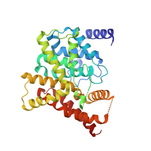
    3B2R

  • PubMed Abstract: 

    Vardenafil has higher affinity to phosphodiesterase-5 (PDE5) than sildenafil and lower administered dosage for the treatment of erectile dysfunction. However, the molecular basis for these differences is puzzling because two drugs have similar chemical structures. Reported here is a crystal structure of the fully active and nonmutated PDE5A1 catalytic domain in complex with vardenafil. The structure shows that the conformation of the H-loop in the PDE5A1-vardenafil complex is different from those of any known structures of the unliganded PDE5 and its complexes with the inhibitors. In addition, the molecular configuration of vardenafil differs from that of sildenafil when bound to PDE5. It is noteworthy that the binding of vardenafil causes loss of the divalent metal ions that have been observed in all the previously published PDE structures. The conformational variation of both PDE5 and the inhibitors provides structural insight into the different potencies of the drugs.


  • Organizational Affiliation
    • Department of Biochemistry and Biophysics, The University of North Carolina, Chapel Hill, NC 27599-7260, USA.

Macromolecule Content 

  • Total Structure Weight: 77.38 kDa 
  • Atom Count: 4,967 
  • Modeled Residue Count: 584 
  • Deposited Residue Count: 660 
  • Unique protein chains: 1

Macromolecules

Find similar proteins by:|  3D Structure
Entity ID: 1
MoleculeChains  Sequence LengthOrganismDetailsImage
cGMP-specific 3',5'-cyclic phosphodiesterase
A, B
330Homo sapiensMutation(s): 0 
Gene Names: PDE5APDE5
EC: 3.1.4.35
UniProt & NIH Common Fund Data Resources
Find proteins for O76074 (Homo sapiens)
Explore O76074 
Go to UniProtKB:  O76074
PHAROS:  O76074
GTEx:  ENSG00000138735 
Entity Groups
Sequence Clusters30% Identity50% Identity70% Identity90% Identity95% Identity100% Identity
UniProt GroupO76074
Sequence Annotations
Expand
Reference Sequence

Small Molecules

Ligands 1 Unique
IDChains Name / Formula / InChI Key2D Diagram3D Interactions
VDN

Query on VDN


Download:Ideal Coordinates CCD File
C [auth A],
D [auth B]
2-{2-ETHOXY-5-[(4-ETHYLPIPERAZIN-1-YL)SULFONYL]PHENYL}-5-METHYL-7-PROPYLIMIDAZO[5,1-F][1,2,4]TRIAZIN-4(1H)-ONE
C23 H32 N6 O4 S
SECKRCOLJRRGGV-UHFFFAOYSA-N
Binding Affinity Annotations 
IDSourceBinding Affinity
VDN BindingDB:  3B2R Ki: 0.7 (nM) from 1 assay(s)
IC50: min: 0.03, max: 0.89 (nM) from 4 assay(s)

Experimental Data & Validation

Experimental Data

  • Method: X-RAY DIFFRACTION
  • Resolution: 2.07 Å
  • R-Value Free:  0.248 (Depositor), 0.247 (DCC) 
  • R-Value Work:  0.216 (Depositor), 0.215 (DCC) 
Space Group: P 21 21 21
Unit Cell:
Length ( Å )Angle ( ˚ )
a = 68.922α = 90
b = 87.822β = 90
c = 138.472γ = 90
Software Package:
Software NamePurpose
ADSCdata collection
AMoREphasing
CNSrefinement
HKL-2000data reduction
HKL-2000data scaling

Structure Validation

View Full Validation Report


Ligand Structure Quality Assessment 


Entry History 

Deposition Data

Revision History  (Full details and data files)

  • Version 1.0: 2008-05-20
    Type: Initial release
  • Version 1.1: 2011-07-13
    Changes: Version format compliance
  • Version 1.2: 2024-02-21
    Changes: Data collection, Database references, Derived calculations
  • Version 1.3: 2024-04-03
    Changes: Refinement description