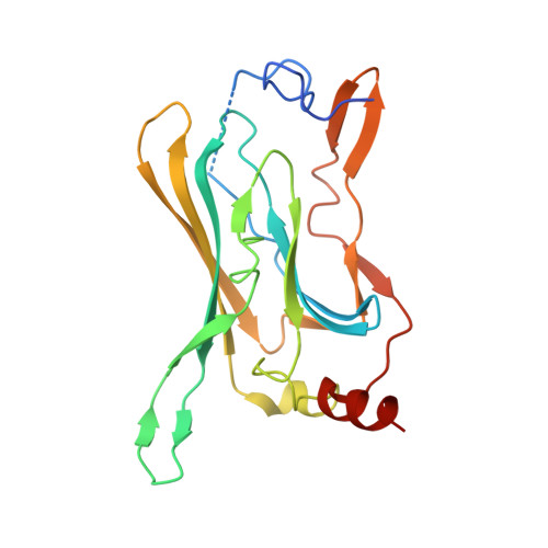
Interactions of cations with the cytoplasmic pores of inward rectifier K(+) channels in the closed state
Inanobe, A., Nakagawa, A., Kurachi, Y.(2011) J Biological Chem 286: 41801-41811
- PubMed: 21982822
- DOI: https://doi.org/10.1074/jbc.M111.278531
- Primary Citation of Related Structures:
3AT8, 3AT9, 3ATA, 3ATB, 3ATD, 3ATE, 3ATF - PubMed Abstract:
Ion channels gate at membrane-embedded domains by changing their conformation along the ion conduction pathway. Inward rectifier K(+) (Kir) channels possess a unique extramembrane cytoplasmic domain that extends this pathway. However, the relevance and contribution of this domain to ion permeation remain unclear. By qualitative x-ray crystallographic analysis, we found that the pore in the cytoplasmic domain of Kir3.2 binds cations in a valency-dependent manner and does not allow the displacement of Mg(2+) by monovalent cations or spermine. Electrophysiological analyses revealed that the cytoplasmic pore of Kir3.2 selectively binds positively charged molecules and has a higher affinity for Mg(2+) when it has a low probability of being open. The selective blocking of chemical modification of the side chain of pore-facing residues by Mg(2+) indicates that the mode of binding of Mg(2+) is likely to be similar to that observed in the crystal structure. These results indicate that the Kir3.2 crystal structure has a closed conformation with a negative electrostatic field potential at the cytoplasmic pore, the potential of which may be controlled by conformational changes in the cytoplasmic domain to regulate ion diffusion along the pore.
- Department of Pharmacology, Graduate School of Medicine, Osaka University, Osaka 565-0871, Japan; Center for Advanced Medical Engineering and Informatics, Osaka University, Osaka 565-0871, Japan. Electronic address: inanobe@pharma2.med.osaka-u.ac.jp.
Organizational Affiliation:


