3ANY

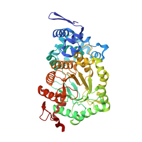
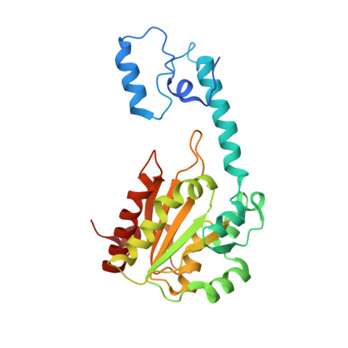
Crystal structure of ethanolamine ammonia-lyase from escherichia coli complexed with CN-CBL and (R)-2-amino-1-propanol


Experimental Data Snapshot

  • Method: X-RAY DIFFRACTION
  • Resolution: 2.10 Å
  • R-Value Free: 0.276 
  • R-Value Work: 0.246 
  • R-Value Observed: 0.248 

Starting Model: experimental
View more details

wwPDB Validation   3D Report Full Report


Ligand Structure Quality Assessment 


This is version 1.3 of the entry. See complete history


Literature

How coenzyme B12-dependent ethanolamine ammonia-lyase deals with both enantiomers of 2-amino-1-propanol as substrates: structure-based rationalization.

Shibata, N.Higuchi, Y.Toraya, T.

(2011) Biochemistry 50: 591-598

  • DOI: https://doi.org/10.1021/bi101696h
  • Primary Citation of Related Structures:  
    3ANY, 3AO0

  • PubMed Abstract: 

    Coenzyme B(12)-dependent ethanolamine ammonia-lyase acts on both enantiomers of the substrate 2-amino-1-propanol [Diziol, P., et al. (1980) Eur. J. Biochem. 106, 211-224]. To rationalize this apparent lack of stereospecificity and the enantiomer-specific stereochemical courses of the deamination, we analyzed the X-ray structures of enantiomer-bound forms of the enzyme-cyanocobalamin complex. The lower affinity for the (R)-enantiomer may be due to the conformational change of the Valα326 side chain of the enzyme. In a manner consistent with the reported experimental results, we can predict that the pro-S hydrogen atom on C1 is abstracted by the adenosyl radical from both enantiomeric substrates, because it is the nearest one in both enantiomer-bound forms. We also predicted that the NH(2) group migrates from C2 to C1 by a suprafacial shift, with inversion of configuration at C1 for both enantiomeric substrates, although the absolute configuration of the 1-amino-1-propanol intermediate is not yet known. Reported labeling experiments demonstrate that (R)-2-amino-1-propanol is deaminated by the enzyme with inversion of configuration at C2, whereas the (S)-enantiomer is deaminated with retention. By taking these results into consideration, we can predict the rotameric radical intermediate from the (S)-enantiomer undergoes flipping to the rotamer from the (R)-enantiomer before the hydrogen back-abstraction. This suggests the preference of the enzyme active site for the rotamer from the (R)-enantiomer in equilibration. This preference might be explained in terms of the steric repulsion of the (S)-enantiomer-derived product radical at C3 with the Pheα329 and Leuα402 residues.


  • Organizational Affiliation

    Department of Life Science, Graduate School of Life Science, University of Hyogo, 3-2-1 Koto, Kamigori-cho, Ako-gun, Hyogo, Japan. shibach@sci.u-hyogo.ac.jp


Macromolecules
Find similar proteins by:  (by identity cutoff)  |  3D Structure
Entity ID: 1
MoleculeChains Sequence LengthOrganismDetailsImage
Ethanolamine ammonia-lyase heavy chain
A, C
453Escherichia coli K-12Mutation(s): 0 
Gene Names: eutBb2441JW2434
EC: 4.3.1.7
UniProt
Find proteins for P0AEJ6 (Escherichia coli (strain K12))
Explore P0AEJ6 
Go to UniProtKB:  P0AEJ6
Entity Groups  
Sequence Clusters30% Identity50% Identity70% Identity90% Identity95% Identity100% Identity
UniProt GroupP0AEJ6
Sequence Annotations
Expand
  • Reference Sequence
Find similar proteins by:  (by identity cutoff)  |  3D Structure
Entity ID: 2
MoleculeChains Sequence LengthOrganismDetailsImage
Ethanolamine ammonia-lyase light chain
B, D
263Escherichia coli K-12Mutation(s): 0 
Gene Names: eutCb2440JW2433
EC: 4.3.1.7
UniProt
Find proteins for P19636 (Escherichia coli (strain K12))
Explore P19636 
Go to UniProtKB:  P19636
Entity Groups  
Sequence Clusters30% Identity50% Identity70% Identity90% Identity95% Identity100% Identity
UniProt GroupP19636
Sequence Annotations
Expand
  • Reference Sequence
Experimental Data & Validation

Experimental Data

  • Method: X-RAY DIFFRACTION
  • Resolution: 2.10 Å
  • R-Value Free: 0.276 
  • R-Value Work: 0.246 
  • R-Value Observed: 0.248 
  • Space Group: P 63
Unit Cell:
Length ( Å )Angle ( ˚ )
a = 244.073α = 90
b = 244.073β = 90
c = 76.98γ = 120
Software Package:
Software NamePurpose
BSSdata collection
REFMACrefinement
HKL-2000data reduction
HKL-2000data scaling

Structure Validation

View Full Validation Report


Ligand Structure Quality Assessment 


Entry History 

Deposition Data

  • Released Date: 2011-08-03 
  • Deposition Author(s): Shibata, N.

Revision History  (Full details and data files)

  • Version 1.0: 2011-08-03
    Type: Initial release
  • Version 1.1: 2012-10-24
    Changes: Non-polymer description
  • Version 1.2: 2017-10-11
    Changes: Refinement description
  • Version 1.3: 2023-11-01
    Changes: Data collection, Database references, Derived calculations, Refinement description