3AMZ

Bovine Xanthine Oxidoreductase urate bound form


Experimental Data Snapshot

  • Method: X-RAY DIFFRACTION
  • Resolution: 2.10 Å
  • R-Value Free: 0.202 
  • R-Value Work: 0.151 
  • R-Value Observed: 0.153 

wwPDB Validation   3D Report Full Report


Ligand Structure Quality Assessment 


This is version 1.2 of the entry. See complete history


Literature

Crystal Structures of Urate Bound Form of Xanthine Oxidoreductase: Substrate Orientation and Structure of the Key Reaction Intermediate

Okamoto, K.Kawaguchi, Y.Eger, B.T.Pai, E.F.Nishino, T.

(2010) J Am Chem Soc 132: 17080-17083

  • DOI: https://doi.org/10.1021/ja1077574
  • Primary Citation of Related Structures:  
    3AMZ, 3AN1

  • PubMed Abstract: 

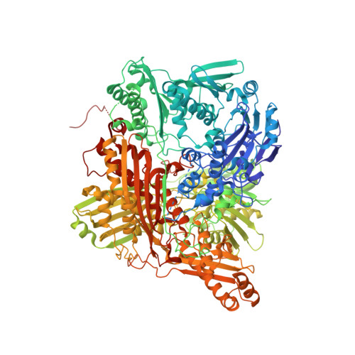
    Two contradictory models have been proposed for the binding mode of the substrate xanthine to and its activation mechanism by xanthine oxidoreductase. In an effort to distinguish between the two models, we determined the crystal structures of the urate complexes of the demolybdo-form of the D428A mutant of rat xanthine oxidoreductase at 1.7 Å and of the reduced bovine milk enzyme at 2.1 Å, the latter representing a reaction intermediate. The results clearly indicate the catalytically relevant binding mode of the substrate xanthine.


  • Organizational Affiliation

    Department of Biochemistry and Molecular Biology, Nippon Medical School, 1-1-5 Sendagi, Bunkyou-ku, Tokyo 113-8602, Japan.


Macromolecules
Find similar proteins by:  (by identity cutoff)  |  3D Structure
Entity ID: 1
MoleculeChains Sequence LengthOrganismDetailsImage
Xanthine dehydrogenase/oxidase
A, B
1,332Bos taurusMutation(s): 0 
EC: 1.17.1.4 (PDB Primary Data), 1.17.3.2 (PDB Primary Data)
UniProt
Find proteins for P80457 (Bos taurus)
Explore P80457 
Go to UniProtKB:  P80457
Entity Groups  
Sequence Clusters30% Identity50% Identity70% Identity90% Identity95% Identity100% Identity
UniProt GroupP80457
Sequence Annotations
Expand
  • Reference Sequence
Small Molecules
Ligands 9 Unique
IDChains Name / Formula / InChI Key2D Diagram3D Interactions
FAD
Query on FAD

Download Ideal Coordinates CCD File 
F [auth A],
Q [auth B]
FLAVIN-ADENINE DINUCLEOTIDE
C27 H33 N9 O15 P2
VWWQXMAJTJZDQX-UYBVJOGSSA-N
NAI
Query on NAI

Download Ideal Coordinates CCD File 
G [auth A],
R [auth B]
1,4-DIHYDRONICOTINAMIDE ADENINE DINUCLEOTIDE
C21 H29 N7 O14 P2
BOPGDPNILDQYTO-NNYOXOHSSA-N
MTE
Query on MTE

Download Ideal Coordinates CCD File 
K [auth A],
U [auth B]
PHOSPHONIC ACIDMONO-(2-AMINO-5,6-DIMERCAPTO-4-OXO-3,7,8A,9,10,10A-HEXAHYDRO-4H-8-OXA-1,3,9,10-TETRAAZA-ANTHRACEN-7-YLMETHYL)ESTER
C10 H14 N5 O6 P S2
HPEUEJRPDGMIMY-IFQPEPLCSA-N
FES
Query on FES

Download Ideal Coordinates CCD File 
C [auth A],
D [auth A],
N [auth B],
O [auth B]
FE2/S2 (INORGANIC) CLUSTER
Fe2 S2
NIXDOXVAJZFRNF-UHFFFAOYSA-N
URC
Query on URC

Download Ideal Coordinates CCD File 
M [auth A],
W [auth B]
URIC ACID
C5 H4 N4 O3
LEHOTFFKMJEONL-UHFFFAOYSA-N
MOS
Query on MOS

Download Ideal Coordinates CCD File 
L [auth A],
V [auth B]
DIOXOTHIOMOLYBDENUM(VI) ION
H Mo O2 S
BDSRWPHSAKXXRG-UHFFFAOYSA-M
GOL
Query on GOL

Download Ideal Coordinates CCD File 
I [auth A],
J [auth A],
T [auth B]
GLYCEROL
C3 H8 O3
PEDCQBHIVMGVHV-UHFFFAOYSA-N
BCT
Query on BCT

Download Ideal Coordinates CCD File 
H [auth A],
S [auth B]
BICARBONATE ION
C H O3
BVKZGUZCCUSVTD-UHFFFAOYSA-M
CA
Query on CA

Download Ideal Coordinates CCD File 
E [auth A],
P [auth B]
CALCIUM ION
Ca
BHPQYMZQTOCNFJ-UHFFFAOYSA-N
Experimental Data & Validation

Experimental Data

  • Method: X-RAY DIFFRACTION
  • Resolution: 2.10 Å
  • R-Value Free: 0.202 
  • R-Value Work: 0.151 
  • R-Value Observed: 0.153 
  • Space Group: C 1 2 1
Unit Cell:
Length ( Å )Angle ( ˚ )
a = 168.211α = 90
b = 124.53β = 91.07
c = 148.025γ = 90
Software Package:
Software NamePurpose
HKL-2000data collection
AMoREphasing
REFMACrefinement
HKL-2000data reduction
HKL-2000data scaling

Structure Validation

View Full Validation Report


Ligand Structure Quality Assessment 


Entry History 

Deposition Data

Revision History  (Full details and data files)

  • Version 1.0: 2010-12-01
    Type: Initial release
  • Version 1.1: 2011-07-13
    Changes: Version format compliance
  • Version 1.2: 2023-11-01
    Changes: Data collection, Database references, Derived calculations, Refinement description