3AKA | pdb_00003aka

Structural basis for prokaryotic calcium-mediated regulation by a Streptomyces coelicolor calcium-binding protein


Experimental Data Snapshot

  • Method: X-RAY DIFFRACTION
  • Resolution: 1.80 Å
  • R-Value Free: 
    0.248 (Depositor), 0.251 (DCC) 
  • R-Value Work: 
    0.197 (Depositor), 0.197 (DCC) 
  • R-Value Observed: 
    0.199 (Depositor) 

Starting Model: experimental
View more details

wwPDB Validation 3D Report Full Report

Validation slider image for 3AKA

This is version 1.3 of the entry. See complete history

Literature

Structural basis for prokaryotic calciummediated regulation by a Streptomyces coelicolor calcium binding protein

Zhao, X.Pang, H.Wang, S.Zhou, W.Yang, K.Bartlam, M.

(2010) Protein Cell 1: 771-779

  • DOI: https://doi.org/10.1007/s13238-010-0085-z
  • Primary Citation Related Structures: 
    3AKA, 3AKB

  • PubMed Abstract: 

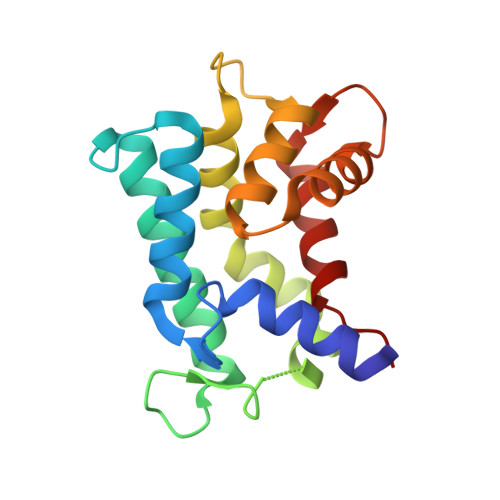
    The important and diverse regulatory roles of Ca(2+) in eukaryotes are conveyed by the EF-hand containing calmodulin superfamily. However, the calcium-regulatory proteins in prokaryotes are still poorly understood. In this study, we report the three-dimensional structure of the calcium-binding protein from Streptomyces coelicolor, named CabD, which shares low sequence homology with other known helix-loop-helix EF-hand proteins. The CabD structure should provide insights into the biological role of the prokaryotic calcium-binding proteins. The unusual structural features of CabD compared with prokaryotic EF-hand proteins and eukaryotic sarcoplasmic calcium-binding proteins, including the bending conformation of the first C-terminal α-helix, unpaired ligand-binding EF-hands and the lack of the extreme C-terminal loop region, suggest it may have a distinct and significant function in calcium-mediated bacterial physiological processes, and provide a structural basis for potential calcium-mediated regulatory roles in prokaryotes.


  • Organizational Affiliation
    • Laboratory of Structural Biology, Tsinghua University, Beijing 100084, China.

Macromolecule Content 

  • Total Structure Weight: 18.03 kDa 
  • Atom Count: 1,443 
  • Modeled Residue Count: 162 
  • Deposited Residue Count: 166 
  • Unique protein chains: 1

Macromolecules

Find similar proteins by:|  3D Structure
Entity ID: 1
MoleculeChains  Sequence LengthOrganismDetailsImage
Putative calcium binding protein166Streptomyces coelicolor A3(2)Mutation(s): 0 
Gene Names: cabdSC6F11.09SCO4411
UniProt
Find proteins for Q9F377 (Streptomyces coelicolor (strain ATCC BAA-471 / A3(2) / M145))
Explore Q9F377 
Go to UniProtKB:  Q9F377
Entity Groups
Sequence Clusters30% Identity50% Identity70% Identity90% Identity95% Identity100% Identity
UniProt GroupQ9F377
Sequence Annotations
Expand
Reference Sequence

Small Molecules

Ligands 1 Unique
IDChains Name / Formula / InChI Key2D Diagram3D Interactions
CA

Query on CA


Download:Ideal Coordinates CCD File
B [auth A]
C [auth A]
D [auth A]
E [auth A]
F [auth A]
B [auth A],
C [auth A],
D [auth A],
E [auth A],
F [auth A],
G [auth A],
H [auth A],
I [auth A],
J [auth A]
CALCIUM ION
Ca
BHPQYMZQTOCNFJ-UHFFFAOYSA-N

Experimental Data & Validation

Experimental Data

  • Method: X-RAY DIFFRACTION
  • Resolution: 1.80 Å
  • R-Value Free:  0.248 (Depositor), 0.251 (DCC) 
  • R-Value Work:  0.197 (Depositor), 0.197 (DCC) 
  • R-Value Observed: 0.199 (Depositor) 
Space Group: P 21 21 21
Unit Cell:
Length ( Å )Angle ( ˚ )
a = 32.876α = 90
b = 51.044β = 90
c = 86.998γ = 90
Software Package:
Software NamePurpose
MAR345dtbdata collection
PHASERphasing
REFMACrefinement
HKL-2000data reduction
HKL-2000data scaling

Structure Validation

View Full Validation Report


Entry History 

Deposition Data

Revision History  (Full details and data files)

  • Version 1.0: 2011-01-26
    Type: Initial release
  • Version 1.1: 2011-07-13
    Changes: Version format compliance
  • Version 1.2: 2024-03-13
    Changes: Data collection, Database references, Derived calculations
  • Version 1.3: 2024-04-03
    Changes: Refinement description