3AHW | pdb_00003ahw

Crystal Structure of Ustilago sphaerogena Ribonuclease U2 complexed with adenosine 2'-monophosphate


Experimental Data Snapshot

  • Method: X-RAY DIFFRACTION
  • Resolution: 1.03 Å
  • R-Value Free: 
    0.144 (Depositor), 0.148 (DCC) 
  • R-Value Work: 
    0.137 (Depositor), 0.144 (DCC) 
  • R-Value Observed: 
    0.137 (Depositor) 

Starting Model: experimental
View more details

wwPDB Validation 3D Report Full Report

Validation slider image for 3AHW

Ligand Structure Quality Assessment 


This is version 1.4 of the entry. See complete history

Literature

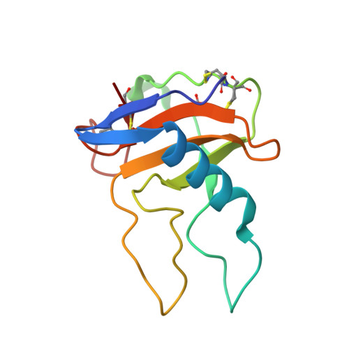
Conformational variation revealed by the crystal structure of RNase U2A complexed with Ca ion and 2'-adenylic acid at 1.03 angstrom resolution.

Noguchi, S.

(2010) Protein Pept Lett 17: 1559-1561

  • DOI: https://doi.org/10.2174/0929866511009011559

  • PubMed Abstract: 

    Asparagine can be non-enzymatically deamidated and isomerized via succinimide to isoaspartate. This post-translational modification can potentially alter the physical properties or the function of the parent protein. Asn32 of ribonuclease U2A from Ustilago sphaerogena is known to rapidly deamidate and isomerize in alkaline conditions. The crystal structure of ribonuclease U2A complexed with 2'-adenylic acid and calcium ions was determined at 1.03 Å resolution. In this structure, the region from Asp29 to Asp37 winds around a calcium ion, and the main-chain of Asn32-Gly33 adopts an extended conformation. Rotation of the side-chain of Asn32 could bring Asn32C(γ) into close proximity to Gly33N, in a conformation suitable for succinimide formation. The structure suggests that in solution the region around Asn32-Gly33 is likely to be in equilibrium between multiple conformers, with the deamidation of Asn32 proceeding when the region adopts an extended conformation.


  • Organizational Affiliation
    • Graduate School of Pharmaceutical Sciences, The University of Tokyo, Tokyo 113-0033, Japan. snoguchi@mol.f.u-tokyo.ac.jp

Macromolecule Content 

  • Total Structure Weight: 12.9 kDa 
  • Atom Count: 1,090 
  • Modeled Residue Count: 114 
  • Deposited Residue Count: 114 
  • Unique protein chains: 1

Macromolecules

Find similar proteins by:|  3D Structure
Entity ID: 1
MoleculeChains  Sequence LengthOrganismDetailsImage
Ribonuclease U2114Ustilago sphaerogenaMutation(s): 0 
EC: 3.1.27.4 (PDB Primary Data), 4.6.1.20 (UniProt)
UniProt
Find proteins for P00654 (Ustilago sphaerogena)
Explore P00654 
Go to UniProtKB:  P00654
Entity Groups
Sequence Clusters30% Identity50% Identity70% Identity90% Identity95% Identity100% Identity
UniProt GroupP00654
Sequence Annotations
Expand
Reference Sequence

Experimental Data & Validation

Experimental Data

  • Method: X-RAY DIFFRACTION
  • Resolution: 1.03 Å
  • R-Value Free:  0.144 (Depositor), 0.148 (DCC) 
  • R-Value Work:  0.137 (Depositor), 0.144 (DCC) 
  • R-Value Observed: 0.137 (Depositor) 
Space Group: P 1 21 1
Unit Cell:
Length ( Å )Angle ( ˚ )
a = 38.994α = 90
b = 35.94β = 103.42
c = 36.151γ = 90
Software Package:
Software NamePurpose
REFMACrefinement
PDB_EXTRACTdata extraction
HKL-2000data collection
HKL-2000data reduction
HKL-2000data scaling
MOLREPphasing

Structure Validation

View Full Validation Report


Ligand Structure Quality Assessment 


Entry History 

Deposition Data

  • Released Date: 2010-07-07 
  • Deposition Author(s): Noguchi, S.

Revision History  (Full details and data files)

  • Version 1.0: 2010-07-07
    Type: Initial release
  • Version 1.1: 2011-07-13
    Changes: Version format compliance
  • Version 1.2: 2019-10-16
    Changes: Data collection, Database references
  • Version 1.3: 2023-11-01
    Changes: Data collection, Database references, Derived calculations, Refinement description
  • Version 1.4: 2024-10-16
    Changes: Structure summary