3AHS | pdb_00003ahs

Crystal Structure of Ustilago sphaerogena Ribonuclease U2B


Experimental Data Snapshot

  • Method: X-RAY DIFFRACTION
  • Resolution: 1.32 Å
  • R-Value Free: 
    0.174 (Depositor), 0.178 (DCC) 
  • R-Value Work: 
    0.162 (Depositor), 0.166 (DCC) 
  • R-Value Observed: 
    0.163 (Depositor) 

Starting Model: experimental
View more details

wwPDB Validation 3D Report Full Report

Validation slider image for 3AHS

This is version 1.4 of the entry. See complete history

Literature

Structural changes induced by the deamidation and isomerization of asparagine revealed by the crystal structure of Ustilago sphaerogena ribonuclease U2B

Noguchi, S.

(2010) Biopolymers 93: 1003-1010

  • DOI: https://doi.org/10.1002/bip.21514
  • Primary Citation Related Structures: 
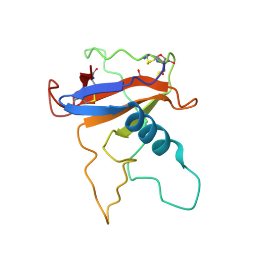
    3AHS

  • PubMed Abstract: 

    Under physiological conditions, the deamidation and isomerization of asparagine to isoaspartate (isoAsp) proceeds nonenzymatically via succinimide. Although a large number of proteins have been reported to contain isoAsp, information concerning the three-dimensional structure of proteins containing isoaspartate is still limited. We have crystallized isoAsp containing Ustilago sphaerogena ribonuclease U2B, and determined the crystal structure at 1.32 Å resolution. The structure revealed that the formation of isoAsp32 induces a single turn unfolding of the α-helix from Asp29 to Asp34, and the region from Asp29 to Arg35 forms a U-shaped loop structure. The electron density map shows that isoAsp32 retained the L-configuration at the C(α) atom. IsoAsp32 is in gauche conformation about a C(α)--C(β) bond, and the polypeptide chain bends by ∼90° at isoAsp32. IsoAsp32 protrudes from the surface of the protein, and the abnormal β-peptide bond in the main-chain and α-carboxylate in the side-chain is fully exposed. The structure suggests that the deamidation of the Asn and the isoAsp formation in proteins could confer immunogenicity.


  • Organizational Affiliation
    • Graduate School of Pharmaceutical Sciences, The University of Tokyo, Bunkyo-Ku, Tokyo 113-0033, Japan. snoguchi@mol.f.u-tokyo.ac.jp

Macromolecule Content 

  • Total Structure Weight: 37.73 kDa 
  • Atom Count: 3,090 
  • Modeled Residue Count: 336 
  • Deposited Residue Count: 342 
  • Unique protein chains: 1

Macromolecules

Find similar proteins by:|  3D Structure
Entity ID: 1
MoleculeChains  Sequence LengthOrganismDetailsImage
Ribonuclease U2
A, B, C
114Ustilago sphaerogenaMutation(s): 0 
EC: 3.1.27.4 (PDB Primary Data), 4.6.1.20 (UniProt)
UniProt
Find proteins for P00654 (Ustilago sphaerogena)
Explore P00654 
Go to UniProtKB:  P00654
Entity Groups
Sequence Clusters30% Identity50% Identity70% Identity90% Identity95% Identity100% Identity
UniProt GroupP00654
Sequence Annotations
Expand
Reference Sequence

Small Molecules

Ligands 3 Unique
IDChains Name / Formula / InChI Key2D Diagram3D Interactions
PO4

Query on PO4


Download:Ideal Coordinates CCD File
D [auth A],
G [auth B],
I [auth C]
PHOSPHATE ION
O4 P
NBIIXXVUZAFLBC-UHFFFAOYSA-K
GOL

Query on GOL


Download:Ideal Coordinates CCD File
F [auth A],
J [auth C]
GLYCEROL
C3 H8 O3
PEDCQBHIVMGVHV-UHFFFAOYSA-N
K

Query on K


Download:Ideal Coordinates CCD File
E [auth A],
H [auth B]
POTASSIUM ION
K
NPYPAHLBTDXSSS-UHFFFAOYSA-N

Experimental Data & Validation

Experimental Data

  • Method: X-RAY DIFFRACTION
  • Resolution: 1.32 Å
  • R-Value Free:  0.174 (Depositor), 0.178 (DCC) 
  • R-Value Work:  0.162 (Depositor), 0.166 (DCC) 
  • R-Value Observed: 0.163 (Depositor) 
Space Group: P 31 2 1
Unit Cell:
Length ( Å )Angle ( ˚ )
a = 98.237α = 90
b = 98.237β = 90
c = 87.605γ = 120
Software Package:
Software NamePurpose
REFMACrefinement
PDB_EXTRACTdata extraction
HKL-2000data reduction
HKL-2000data scaling
MOLREPphasing

Structure Validation

View Full Validation Report


Entry History 

Deposition Data

  • Released Date: 2010-07-07 
  • Deposition Author(s): Noguchi, S.

Revision History  (Full details and data files)

  • Version 1.0: 2010-07-07
    Type: Initial release
  • Version 1.1: 2011-07-13
    Changes: Version format compliance
  • Version 1.2: 2011-07-27
    Changes: Database references, Derived calculations, Non-polymer description
  • Version 1.3: 2023-11-01
    Changes: Advisory, Data collection, Database references, Derived calculations, Refinement description
  • Version 1.4: 2024-10-23
    Changes: Structure summary