3AGH | pdb_00003agh

X-ray analysis of lysozyme in the presence of 200 mM Arg


Experimental Data Snapshot

  • Method: X-RAY DIFFRACTION
  • Resolution: 1.49 Å
  • R-Value Free: 
    0.205 (Depositor), 0.203 (DCC) 
  • R-Value Work: 
    0.175 (Depositor), 0.175 (DCC) 
  • R-Value Observed: 
    0.176 (Depositor) 

Starting Model: experimental
View more details

wwPDB Validation 3D Report Full Report

Validation slider image for 3AGH

This is version 1.3 of the entry. See complete history

Literature

High-resolution X-ray analysis reveals binding of arginine to aromatic residues of lysozyme surface: implication of suppression of protein aggregation by arginine

Ito, L.Shiraki, K.Matsuura, T.Okumura, M.Hasegawa, K.Baba, S.Yamaguchi, H.Kumasaka, T.

(2011) Protein Eng Des Sel 24: 269-274

  • DOI: https://doi.org/10.1093/protein/gzq101
  • Primary Citation Related Structures: 
    3AGG, 3AGH, 3AGI

  • PubMed Abstract: 

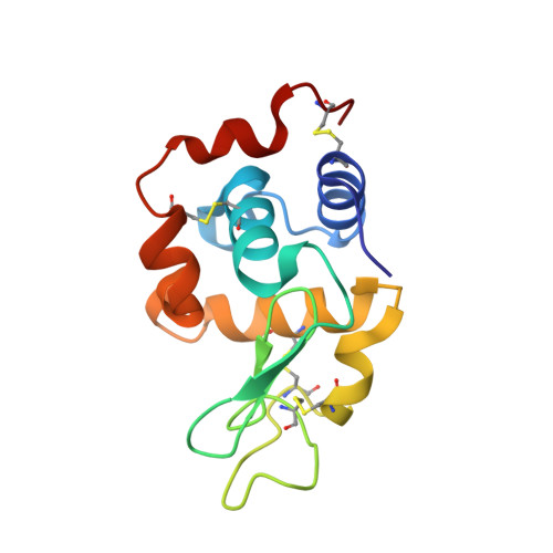
    While biotechnological applications of arginine (Arg) as a solution additive that prevents protein aggregation are increasing, the molecular mechanism of its effects remains unclear. In this study, we investigated the Arg-lysozyme complex by high-resolution crystallographic analysis. Three Arg molecules were observed to be in close proximity to aromatic amino acid residues of the protein surface, and their occupancies gradually increased with increasing Arg concentration. These interactions were mediated by electrostatic, hydrophobic and cation-π interactions with the surface residues. The binding of Arg decreased the accessible surface area of aromatic residues by 40%, but increased that of charged residues by 10%. These changes might prevent intermolecular hydrophobic interactions by shielding hydrophobic regions of the lysozyme surface, resulting in an increase in protein solubility.


  • Organizational Affiliation
    • Japan Synchrotron Radiation Research Institute (SPring-8), 1-1-1 Kouto, Sayo, Hyogo 679-5198, Japan. l-ito@spring8.or.jp

Macromolecule Content 

  • Total Structure Weight: 14.56 kDa 
  • Atom Count: 1,250 
  • Modeled Residue Count: 129 
  • Deposited Residue Count: 129 
  • Unique protein chains: 1

Macromolecules

Find similar proteins by:|  3D Structure
Entity ID: 1
MoleculeChains  Sequence LengthOrganismDetailsImage
Lysozyme C129Gallus gallusMutation(s): 0 
EC: 3.2.1.17
UniProt
Find proteins for P00698 (Gallus gallus)
Explore P00698 
Go to UniProtKB:  P00698
Entity Groups
Sequence Clusters30% Identity50% Identity70% Identity90% Identity95% Identity100% Identity
UniProt GroupP00698
Sequence Annotations
Expand
Reference Sequence

Experimental Data & Validation

Experimental Data

  • Method: X-RAY DIFFRACTION
  • Resolution: 1.49 Å
  • R-Value Free:  0.205 (Depositor), 0.203 (DCC) 
  • R-Value Work:  0.175 (Depositor), 0.175 (DCC) 
  • R-Value Observed: 0.176 (Depositor) 
Space Group: P 43 21 2
Unit Cell:
Length ( Å )Angle ( ˚ )
a = 78.299α = 90
b = 78.299β = 90
c = 37.284γ = 90
Software Package:
Software NamePurpose
HKL-2000data collection
MOLREPphasing
REFMACrefinement
HKL-2000data reduction
HKL-2000data scaling

Structure Validation

View Full Validation Report


Entry History 

Deposition Data

Revision History  (Full details and data files)

  • Version 1.0: 2011-03-23
    Type: Initial release
  • Version 1.1: 2011-07-13
    Changes: Version format compliance
  • Version 1.2: 2023-11-01
    Changes: Data collection, Database references, Derived calculations, Refinement description
  • Version 1.3: 2024-10-30
    Changes: Structure summary