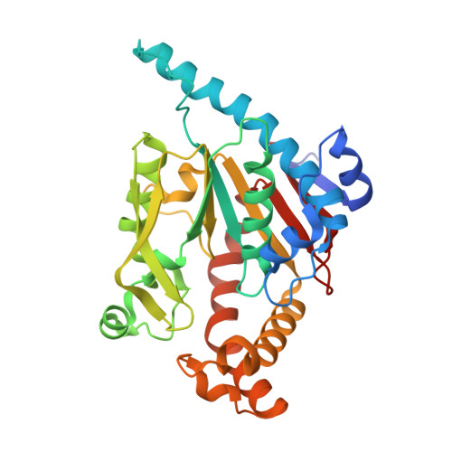
3AF1

Pantothenate kinase from Mycobacterium tuberculosis (MtPanK) in complex with GDP


Experimental Data Snapshot

  • Method: X-RAY DIFFRACTION
  • Resolution: 2.50 Å
  • R-Value Free: 0.243 
  • R-Value Work: 0.193 
  • R-Value Observed: 0.196 

Starting Model: experimental
View more details

wwPDB Validation   3D Report Full Report


Ligand Structure Quality Assessment 


This is version 1.3 of the entry. See complete history


Literature

M. tuberculosis pantothenate kinase: dual substrate specificity and unusual changes in ligand locations

Chetnani, B.Kumar, P.Surolia, A.Vijayan, M.

(2010) J Mol Biol 400: 171-185

  • DOI: https://doi.org/10.1016/j.jmb.2010.04.064
  • Primary Citation of Related Structures:  
    3AEZ, 3AF0, 3AF1, 3AF2, 3AF3, 3AF4

  • PubMed Abstract: 

    Kinetic measurements of enzyme activity indicate that type I pantothenate kinase from Mycobacterium tuberculosis has dual substrate specificity for ATP and GTP, unlike the enzyme from Escherichia coli, which shows a higher specificity for ATP. A molecular explanation for the difference in the specificities of the two homologous enzymes is provided by the crystal structures of the complexes of the M. tuberculosis enzyme with (1) GMPPCP and pantothenate, (2) GDP and phosphopantothenate, (3) GDP, (4) GDP and pantothenate, (5) AMPPCP, and (6) GMPPCP, reported here, and the structures of the complexes of the two enzymes involving coenzyme A and different adenyl nucleotides reported earlier. The explanation is substantially based on two critical substitutions in the amino acid sequence and the local conformational change resulting from them. The structures also provide a rationale for the movement of ligands during the action of the mycobacterial enzyme. Dual specificity of the type exhibited by this enzyme is rare. The change in locations of ligands during action, observed in the case of the M. tuberculosis enzyme, is unusual, so is the striking difference between two homologous enzymes in the geometry of the binding site, locations of ligands, and specificity. Furthermore, the dual specificity of the mycobacterial enzyme appears to have been caused by a biological necessity.


  • Organizational Affiliation

    Molecular Biophysics Unit, Indian Institute of Science, Bangalore 560012, India.


Macromolecules
Find similar proteins by:  (by identity cutoff)  |  3D Structure
Entity ID: 1
MoleculeChains Sequence LengthOrganismDetailsImage
Pantothenate kinase312Mycobacterium tuberculosis H37RvMutation(s): 0 
Gene Names: coaARv1092c
EC: 2.7.1.33
UniProt
Find proteins for P9WPA7 (Mycobacterium tuberculosis (strain ATCC 25618 / H37Rv))
Explore P9WPA7 
Go to UniProtKB:  P9WPA7
Entity Groups  
Sequence Clusters30% Identity50% Identity70% Identity90% Identity95% Identity100% Identity
UniProt GroupP9WPA7
Sequence Annotations
Expand
  • Reference Sequence
Small Molecules
Ligands 4 Unique
IDChains Name / Formula / InChI Key2D Diagram3D Interactions
GDP
Query on GDP

Download Ideal Coordinates CCD File 
B [auth A]GUANOSINE-5'-DIPHOSPHATE
C10 H15 N5 O11 P2
QGWNDRXFNXRZMB-UUOKFMHZSA-N
FLC
Query on FLC

Download Ideal Coordinates CCD File 
C [auth A]CITRATE ANION
C6 H5 O7
KRKNYBCHXYNGOX-UHFFFAOYSA-K
GOL
Query on GOL

Download Ideal Coordinates CCD File 
G [auth A]
H [auth A]
I [auth A]
J [auth A]
K [auth A]
G [auth A],
H [auth A],
I [auth A],
J [auth A],
K [auth A],
L [auth A],
M [auth A],
N [auth A],
O [auth A],
P [auth A],
Q [auth A],
R [auth A],
S [auth A]
GLYCEROL
C3 H8 O3
PEDCQBHIVMGVHV-UHFFFAOYSA-N
CL
Query on CL

Download Ideal Coordinates CCD File 
D [auth A],
E [auth A],
F [auth A]
CHLORIDE ION
Cl
VEXZGXHMUGYJMC-UHFFFAOYSA-M
Experimental Data & Validation

Experimental Data

  • Method: X-RAY DIFFRACTION
  • Resolution: 2.50 Å
  • R-Value Free: 0.243 
  • R-Value Work: 0.193 
  • R-Value Observed: 0.196 
  • Space Group: P 31 2 1
Unit Cell:
Length ( Å )Angle ( ˚ )
a = 103.9α = 90
b = 103.9β = 90
c = 90.21γ = 120
Software Package:
Software NamePurpose
MAR345dtbdata collection
PHASERphasing
REFMACrefinement
MOSFLMdata reduction
SCALAdata scaling

Structure Validation

View Full Validation Report


Ligand Structure Quality Assessment 


Entry History 

Deposition Data

Revision History  (Full details and data files)

  • Version 1.0: 2010-05-26
    Type: Initial release
  • Version 1.1: 2011-07-13
    Changes: Version format compliance
  • Version 1.2: 2014-01-29
    Changes: Database references
  • Version 1.3: 2023-11-01
    Changes: Data collection, Database references, Derived calculations, Refinement description