3ADJ | pdb_00003adj

Structure of Arabidopsis HYL1 and its molecular implications for miRNA processing


Experimental Data Snapshot

  • Method: X-RAY DIFFRACTION
  • Resolution: 3.00 Å
  • R-Value Free: 
    0.322 (Depositor), 0.311 (DCC) 
  • R-Value Work: 
    0.255 (Depositor), 0.253 (DCC) 
  • R-Value Observed: 
    0.261 (Depositor) 

Starting Model: experimental
View more details

wwPDB Validation 3D Report Full Report

Validation slider image for 3ADJ

This is version 1.2 of the entry. See complete history

Literature

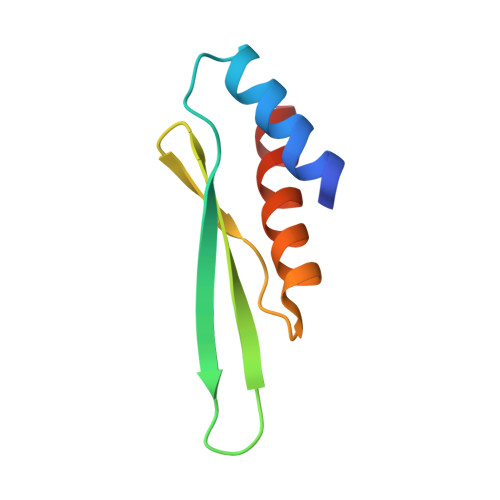
Structure of arabidopsis HYPONASTIC LEAVES1 and its molecular implications for miRNA processing

Yang, S.W.Chen, H.Y.Yang, J.Machida, S.Chua, N.H.Yuan, Y.A.

(2010) Structure 18: 594-605

  • DOI: https://doi.org/10.1016/j.str.2010.02.006
  • Primary Citation Related Structures: 
    3ADG, 3ADI, 3ADJ, 3ADL

  • PubMed Abstract: 

    The Arabidopsis HYPONASTIC LEAVES1 (HYL1) is a double-stranded RNA-binding protein that forms a complex with DICER-LIKE1 (DCL1) and SERRATE to facilitate processing of primary miRNAs into microRNAs (miRNAs). However, the structural mechanisms of miRNA maturation by this complex are poorly understood. Here, we present the crystal structures of double-stranded RNA binding domains (dsRBD1 and dsRBD2) of HYL1 and HYL1 dsRBD1 (HR1)/dsRNA complex as well as human TRBP2 dsRBD2 (TR2)/dsRNA complex for comparison analysis. Structural and functional study demonstrates that both HR1 and TR2 are canonical dsRBDs for dsRNA binding, whereas HR2 of HYL1 is a non-canonical dsRBD harboring a putative dimerization interface. Domain swapping within the context of HYL1 demonstrates that TR2 can supplant the function of HR1 in vitro and in vivo. Further biochemical analyses suggest that HYL1 probably binds to the miRNA/miRNA( *) region of precursors as a dimer mediated by HR2.


  • Organizational Affiliation
    • Host-Pathogen Interaction Group, Temasek Life Sciences Laboratory, National University of Singapore, 1 Research Link, Singapore 117604, Singapore.

Macromolecule Content 

  • Total Structure Weight: 8.28 kDa 
  • Atom Count: 552 
  • Modeled Residue Count: 72 
  • Deposited Residue Count: 76 
  • Unique protein chains: 1

Macromolecules

Find similar proteins by:|  3D Structure
Entity ID: 1
MoleculeChains  Sequence LengthOrganismDetailsImage
F21M12.9 protein76Arabidopsis thalianaMutation(s): 0 
UniProt
Find proteins for O04492 (Arabidopsis thaliana)
Explore O04492 
Go to UniProtKB:  O04492
Entity Groups
Sequence Clusters30% Identity50% Identity70% Identity90% Identity95% Identity100% Identity
UniProt GroupO04492
Sequence Annotations
Expand
Reference Sequence

Experimental Data & Validation

Experimental Data

  • Method: X-RAY DIFFRACTION
  • Resolution: 3.00 Å
  • R-Value Free:  0.322 (Depositor), 0.311 (DCC) 
  • R-Value Work:  0.255 (Depositor), 0.253 (DCC) 
  • R-Value Observed: 0.261 (Depositor) 
Space Group: P 43
Unit Cell:
Length ( Å )Angle ( ˚ )
a = 46.108α = 90
b = 46.108β = 90
c = 33.318γ = 90
Software Package:
Software NamePurpose
HKL-2000data collection
MOLREPphasing
REFMACrefinement
HKL-2000data reduction
HKL-2000data scaling

Structure Validation

View Full Validation Report


Entry History 

Deposition Data

Revision History  (Full details and data files)

  • Version 1.0: 2010-05-26
    Type: Initial release
  • Version 1.1: 2011-07-13
    Changes: Advisory, Version format compliance
  • Version 1.2: 2023-11-01
    Changes: Data collection, Database references, Refinement description