3A5P | pdb_00003a5p

Crystal structure of hemagglutinin


Experimental Data Snapshot

  • Method: X-RAY DIFFRACTION
  • Resolution: 1.82 Å
  • R-Value Free: 
    0.194 (Depositor), 0.193 (DCC) 
  • R-Value Work: 
    0.154 (Depositor), 0.154 (DCC) 
  • R-Value Observed: 
    0.156 (Depositor) 

Starting Model: experimental
View more details

wwPDB Validation 3D Report Full Report

Validation slider image for 3A5P

This is version 1.4 of the entry. See complete history

Literature

The Structure of Physarum polycephalum hemagglutinin I suggests a minimal carbohydrate recognition domain of legume lectin fold

Kouno, T.Watanabe, N.Sakai, N.Nakamura, T.Nabeshima, Y.Morita, M.Mizuguchi, M.Aizawa, T.Demura, M.Imanaka, T.Tanaka, I.Kawano, K.

(2011) J Mol Biology 405: 560-569

  • DOI: https://doi.org/10.1016/j.jmb.2010.11.024
  • Primary Citation Related Structures: 
    3A5P

  • PubMed Abstract: 

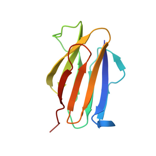
    Physarum polycephalum hemagglutinin I (HA1) is a 104-residue protein that is secreted to extracellular space. The crystal structure of HA1 has a β-sandwich fold found among lectin structures, such as legume lectins and galectins. Interestingly, the β-sandwich of HA1 lacks a jelly roll motif and is essentially composed of two simple up-and-down β-sheets. This up-and-down β-sheet motif is well conserved in other legume lectin-like proteins derived from animals, plants, bacteria, and viruses. It is more noteworthy that the up-and-down β-sheet motif includes many residues that make contact with the target carbohydrates. Our NMR data demonstrate that HA1 lacking a jelly roll motif also binds to its target glycopeptide. Taken together, these data show that the up-and-down β-sheet motif provides a fundamental scaffold for the binding of legume lectin-like proteins to the target carbohydrates, and the structure of HA1 suggests a minimal carbohydrate recognition domain.


  • Organizational Affiliation
    • Department of Structural Biology, University of Toyama, Toyama 930-0194, Japan. konox005@umn.edu

Macromolecule Content 

  • Total Structure Weight: 43.6 kDa 
  • Atom Count: 3,996 
  • Modeled Residue Count: 412 
  • Deposited Residue Count: 416 
  • Unique protein chains: 1

Macromolecules

Find similar proteins by:|  3D Structure
Entity ID: 1
MoleculeChains  Sequence LengthOrganismDetailsImage
Haemagglutinin I
A, B, C, D
104Badhamia polycephalaMutation(s): 0 
UniProt
Find proteins for O60949 (Physarum polycephalum)
Explore O60949 
Go to UniProtKB:  O60949
Entity Groups
Sequence Clusters30% Identity50% Identity70% Identity90% Identity95% Identity100% Identity
UniProt GroupO60949
Sequence Annotations
Expand
Reference Sequence

Experimental Data & Validation

Experimental Data

  • Method: X-RAY DIFFRACTION
  • Resolution: 1.82 Å
  • R-Value Free:  0.194 (Depositor), 0.193 (DCC) 
  • R-Value Work:  0.154 (Depositor), 0.154 (DCC) 
  • R-Value Observed: 0.156 (Depositor) 
Space Group: P 21 21 21
Unit Cell:
Length ( Å )Angle ( ˚ )
a = 64.929α = 90
b = 77.98β = 90
c = 86.109γ = 90
Software Package:
Software NamePurpose
REFMACrefinement
PDB_EXTRACTdata extraction
CrystalCleardata collection
HKL-2000data reduction
HKL-2000data scaling
MOLREPphasing

Structure Validation

View Full Validation Report


Entry History 

Deposition Data

Revision History  (Full details and data files)

  • Version 1.0: 2010-08-11
    Type: Initial release
  • Version 1.1: 2011-07-13
    Changes: Version format compliance
  • Version 1.2: 2014-01-22
    Changes: Database references
  • Version 1.3: 2024-03-13
    Changes: Data collection, Database references
  • Version 1.4: 2024-04-03
    Changes: Refinement description