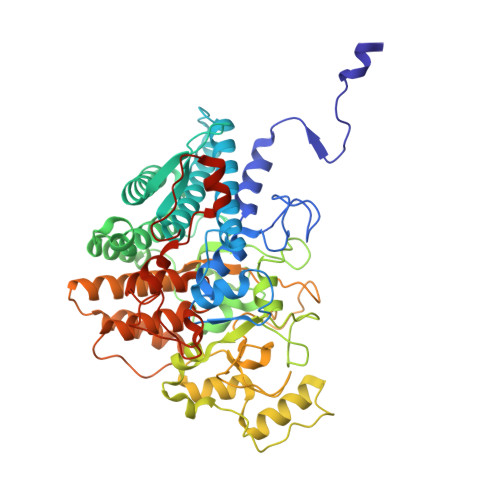
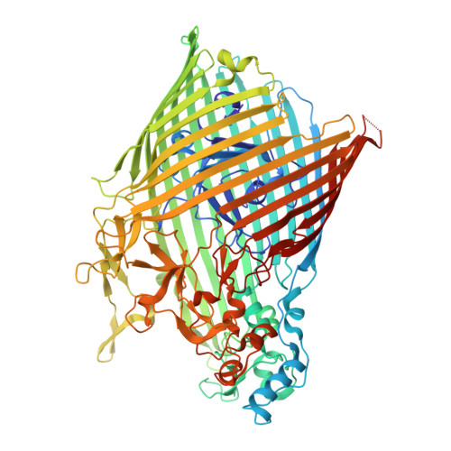
38FE | pdb_000038fe

Bacteroides SusCD + Maltoheptaose - Closed w/ Plug Domain


Experimental Data Snapshot

  • Method: ELECTRON MICROSCOPY
  • Resolution: 2.30 Å
  • Aggregation State: PARTICLE 
  • Reconstruction Method: SINGLE PARTICLE 

wwPDB Validation 3D Report Full Report

Validation slider image for 38FE

This is version 1.0 of the entry. See complete history. 

Literature

Structural analyses of a heterodimeric SusCD complex captures intermediate states of maltooligosaccharide transport.

Cadigan, M.C., Fuentes, N.R., Salmen, W., Roberts, J.R., Ruotolo, B., Ohi, M.D., Koropatkin, N.M.

(2026) J Biol Chem : 113672-113672

  • DOI: https://doi.org/10.1016/j.jbc.2026.113672
  • Primary Citation Related Structures: 
    38AQ, 38FE, 38FH, 38FI

  • PubMed Abstract: 

    Bacteroides are abundant gut bacteria with diverse polysaccharide metabolizing capabilities encoded by co-transcribed polysaccharide utilization loci (PULs). The starch utilization system (Sus) is required for starch and α-glucan metabolism in Bacteroides thetaiotaomicron and has served as a model for PUL studies. However, the mechanism of maltooligosaccharide (MOS) transport and the architecture of the SusCD complex remain incompletely understood. Here, we used cryo-electron microscopy to capture multiple transport-relevant conformations of the SusD lipoprotein and the TonB-dependent transporter SusC in both unliganded and ligand-bound states. In the absence of ligand, SusCD adopts three conformations including an open state in which SusD is displaced from SusC, a state containing the SusC plug, and a state lacking density for the SusC plug. Three-dimensional variability analysis of the structure where SusD is displaced from SusC reveals continuous SusD mobility coupled to movement of the SusC N-terminus. The maltoheptaose-bound SusCD structure reveals two binding sites, one at the SusD-SusC interface and a second within SusC, indicating how MOS stabilizes a closed SusCD assembly. Notably, all structures show that SusCD is a heterodimer, unlike other Sus-like transporters that assemble into SusCD heterotetramers. Native mass spectrometry confirms this heterodimeric stoichiometry, and comparisons with other SusCD-like complexes suggest a structural basis for this distinct assembly of SusCD. Together, these data define substrate binding and intermediate transport states in a canonical PUL system and reveal architectural divergence among Sus-like carbohydrate transport complexes in Bacteroides.


  • Organizational Affiliation: 
    • Department of Microbiology and Immunology, University of Michigan Medical School, Ann Arbor, MI 48109.

Macromolecule Content 

  • Total Structure Weight: 163.48 kDa 
  • Atom Count: 11,402 
  • Modeled Residue Count: 1,420 
  • Deposited Residue Count: 1,433 
  • Unique protein chains: 2

Macromolecules

Find similar proteins by:|  3D Structure
Entity ID: 1
MoleculeChains  Sequence LengthOrganismDetailsImage
Starch-binding protein SusDA [auth B]536Bacteroides thetaiotaomicron VPI-5482Mutation(s): 0 
Gene Names: susD, BT_3701
UniProt
Find proteins for Q8A1G2 (Bacteroides thetaiotaomicron (strain ATCC 29148 / DSM 2079 / JCM 5827 / CCUG 10774 / NCTC 10582 / VPI-5482 / E50))
Explore Q8A1G2 
Go to UniProtKB:  Q8A1G2
Entity Groups
Sequence Clusters30% Identity50% Identity70% Identity90% Identity95% Identity100% Identity
UniProt GroupQ8A1G2
Sequence Annotations
Expand
Reference Sequence
Find similar proteins by:|  3D Structure
Entity ID: 2
MoleculeChains  Sequence LengthOrganismDetailsImage
TonB-dependent receptor SusCB [auth A]897Bacteroides thetaiotaomicron VPI-5482Mutation(s): 0 
Gene Names: susC, BT_3702
UniProt
Find proteins for Q8A1G1 (Bacteroides thetaiotaomicron (strain ATCC 29148 / DSM 2079 / JCM 5827 / CCUG 10774 / NCTC 10582 / VPI-5482 / E50))
Explore Q8A1G1 
Go to UniProtKB:  Q8A1G1
Entity Groups
Sequence Clusters30% Identity50% Identity70% Identity90% Identity95% Identity100% Identity
UniProt GroupQ8A1G1
Sequence Annotations
Expand
Reference Sequence

Oligosaccharides

Help  
Entity ID: 3
MoleculeChains Length2D Diagram GlycosylationD Interactions
alpha-D-glucopyranose-(1-4)-alpha-D-glucopyranose-(1-4)-alpha-D-glucopyranose-(1-4)-alpha-D-glucopyranose-(1-4)-alpha-D-glucopyranose-(1-4)-alpha-D-glucopyranose-(1-4)-beta-D-glucopyranoseC [auth D]7N/A
Entity ID: 4
MoleculeChains Length2D Diagram GlycosylationD Interactions
alpha-D-glucopyranose-(1-4)-alpha-D-glucopyranose-(1-4)-alpha-D-glucopyranose-(1-4)-alpha-D-glucopyranose-(1-4)-alpha-D-glucopyranose-(1-4)-alpha-D-glucopyranose-(1-4)-alpha-D-glucopyranoseD [auth C]7N/A

Experimental Data & Validation

Experimental Data

  • Method: ELECTRON MICROSCOPY
  • Resolution: 2.30 Å
  • Aggregation State: PARTICLE 
  • Reconstruction Method: SINGLE PARTICLE 
EM Software:
TaskSoftware PackageVersion
MODEL REFINEMENTPHENIX2.0_5793
RECONSTRUCTIONcryoSPARC4.0+

Structure Validation

View Full Validation Report


Entry History 

& Funding Information

Deposition Data


Funding OrganizationLocationGrant Number
Not funded--

Revision History  (Full details and data files)

  • Version 1.0: 2026-11-04
    Type: Initial release