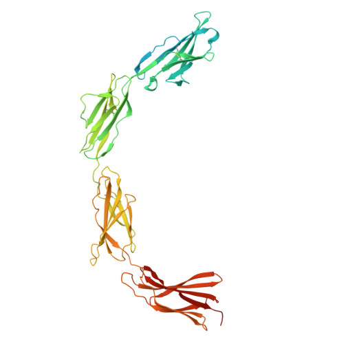
37NT | pdb_000037nt

Crystal structure of the extracellular region of the human receptor tyrosine kinase MERTK


Experimental Data Snapshot

  • Method: X-RAY DIFFRACTION
  • Resolution: 3.41 Å
  • R-Value Free: 
    0.307 (Depositor), 0.308 (DCC) 
  • R-Value Work: 
    0.256 (Depositor), 0.256 (DCC) 
  • R-Value Observed: 
    0.259 (Depositor) 

Starting Model: in silico
View more details

wwPDB Validation 3D Report Full Report

Validation slider image for 37NT

Ligand Structure Quality Assessment 


This is version 1.0 of the entry. See complete history. 

Literature

Structural organization of TAM receptor ectodomains.

Starbird, C.A., Zuo, Y., Walker, K., Stayrook, S.E., Ferguson, K.M.

(2026) Biochem J 

  • DOI: https://doi.org/10.1042/BCJ20260584
  • Primary Citation Related Structures: 
    37NT

  • PubMed Abstract: 

    TYRO3, AXL, and MERTK, the TAM receptor tyrosine kinases (RTKs), play roles in phagocytosis and negative regulation of immune responses, contributing to homeostasis in many adult tissues. Aberrant TAM signaling leads to diseases including autoimmune disorders, viral infection, inflammation and cancer. Development of successful drugs targeting TAM receptors is hindered by incomplete knowledge of the receptor activation mechanism. Receptor dimerization is assumed to play a role, as seen for most RTKs. However, full biological responses require the TAM ligands to engage phosphatidylserine in an opposing cell or virus membrane, suggesting a more complex mechanism, possibly involving higher-order receptor oligomerization or clustering. Here, we present an X ray crystal structure of the ectodomain of MERTK, which adopts a highly curved conformation with key interdomain boundaries stabilized by N-linked glycan moieties. We show that this MERTK extracellular region (ECR) forms a similar conformation in solution using small-angle X ray scattering (SAXS), which also suggests a flexible ECR. We compare these crystallographic and solution data for MERTK with SAXS and AlphaFold structural predictions for the other two TAM receptors. SAXS data confirm that each TAM ECR has a distinct curvature, with TYRO3 the most bent and AXL relatively straight. This leads us to speculate that these different ectodomain conformations guide receptor-specific functions. The MERTK X-ray crystal structure also suggests a head-to-tail intermolecular interaction between the Ig2 domain of one MERTK and the FNIIIb domain of a second that has intriguing parallels with interactions that have been proposed to regulate Eph receptor clustering.


  • Organizational Affiliation: 
    • Yale University School of Medicine, New Haven, Connecticut, United States.

Macromolecule Content 

  • Total Structure Weight: 59.88 kDa 
  • Atom Count: 3,435 
  • Modeled Residue Count: 392 
  • Deposited Residue Count: 490 
  • Unique protein chains: 1

Macromolecules

Find similar proteins by:|  3D Structure
Entity ID: 1
MoleculeChains  Sequence LengthOrganismDetailsImage
Tyrosine-protein kinase Mer490Homo sapiensMutation(s): 0 
Gene Names: MERTK, MER
EC: 2.7.10.1
UniProt & NIH Common Fund Data Resources
Find proteins for Q12866 (Homo sapiens)
Explore Q12866 
Go to UniProtKB:  Q12866
PHAROS:  Q12866
GTEx:  ENSG00000153208 
Entity Groups
Sequence Clusters30% Identity50% Identity70% Identity90% Identity95% Identity100% Identity
UniProt GroupQ12866
Glycosylation
Glycosylation Sites: 10Go to GlyGen: Q12866-1
Sequence Annotations
Expand
Reference Sequence

Oligosaccharides

Help  
Entity ID: 2
MoleculeChains Length2D Diagram GlycosylationD Interactions
beta-D-mannopyranose-(1-4)-2-acetamido-2-deoxy-beta-D-glucopyranose-(1-4)-2-acetamido-2-deoxy-beta-D-glucopyranose
B, C, E
3N-Glycosylation
Entity ID: 3
MoleculeChains Length2D Diagram GlycosylationD Interactions
alpha-D-mannopyranose-(1-3)-[alpha-D-mannopyranose-(1-6)]beta-D-mannopyranose-(1-4)-2-acetamido-2-deoxy-beta-D-glucopyranose-(1-4)-2-acetamido-2-deoxy-beta-D-glucopyranose
D, F, G
5N-Glycosylation
Entity ID: 4
MoleculeChains Length2D Diagram GlycosylationD Interactions
2-acetamido-2-deoxy-beta-D-glucopyranose-(1-4)-2-acetamido-2-deoxy-beta-D-glucopyranose
H, I
2N-Glycosylation
Entity ID: 5
MoleculeChains Length2D Diagram GlycosylationD Interactions
alpha-D-mannopyranose-(1-3)-beta-D-mannopyranose-(1-4)-2-acetamido-2-deoxy-beta-D-glucopyranose-(1-4)-2-acetamido-2-deoxy-beta-D-glucopyranose
J
4N-Glycosylation

Small Molecules

Ligands 1 Unique
IDChains Name / Formula / InChI Key2D Diagram3D Interactions
NAG

Query on NAG


Download:Ideal Coordinates CCD File
K [auth A]2-acetamido-2-deoxy-beta-D-glucopyranose
C8 H15 N O6
OVRNDRQMDRJTHS-FMDGEEDCSA-N

Experimental Data & Validation

Experimental Data

  • Method: X-RAY DIFFRACTION
  • Resolution: 3.41 Å
  • R-Value Free:  0.307 (Depositor), 0.308 (DCC) 
  • R-Value Work:  0.256 (Depositor), 0.256 (DCC) 
  • R-Value Observed: 0.259 (Depositor) 
Space Group: P 21 21 21
Unit Cell:
Length ( Å )Angle ( ˚ )
a = 40.5α = 90
b = 133.514β = 90
c = 163.811γ = 90
Software Package:
Software NamePurpose
PHENIXrefinement
XDSdata reduction
XDSdata scaling
PHASERphasing

Structure Validation

View Full Validation Report


Ligand Structure Quality Assessment 


Entry History 

& Funding Information

Deposition Data


Funding OrganizationLocationGrant Number
National Institutes of Health/National Cancer Institute (NIH/NCI)United StatesCA214704
National Institutes of Health/National Institute of General Medical Sciences (NIH/NIGMS)United StatesGM149406
National Institutes of Health/National Institute of General Medical Sciences (NIH/NIGMS)United StatesGM121460
National Institutes of Health/National Institute of General Medical Sciences (NIH/NIGMS)United StatesGM144683

Revision History  (Full details and data files)

  • Version 1.0: 2026-10-07
    Type: Initial release